
Fig. 2
- ID
- ZDB-FIG-090415-17
- Publication
- Warga et al., 1998 - Spadetail-dependent cell compaction of the dorsal zebrafish blastula
- Other Figures
- All Figure Page
- Back to All Figure Page
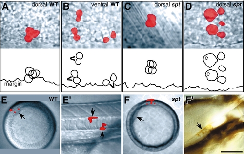
Cell clustering and dorsoventral position of prospective axial mesendoderm is altered in spadetail mutants. (A–D) Labeled clones at 40% epiboly. The upper panel is the record, and the lower panel is a tracing of the individual deep cells, typically by this time there were 4 deep cells descended from the mid-blastula marginal cell (3.7 cells ± 0.22, wild type; 3.9 cells ± 0.39, spadetail mutant); blastoderm margin is indicated; enveloping layer cells (e); recent cell divisions (v’s). Dorsal clones in mutant embryos disperse significantly more than in normal embryos (χ2 = 21.1, df = 4; P < 0.01). (E and F) Labeled clones contributing to notochord in the trunk. (E and F) Animal pole views at shield stage; dorsal side (arrow). (E′ and F′) Side views. (E′) 1 day wildtype embryo. The differentiated clone (arrows), located midtrunk, is notochord sheath, a derivative of the notochord (Melby et al., 1996). (F′) 4 day spadetail-mutant embryo, after whole-mount staining for the fixable tracer. The differentiated clone, now stained brown (arrow), is notochord sheath located in the anterior trunk. Scale bar: 50 μm (A–D), 100 μm (E′ and F′), 250 μm (E and F). |
Reprinted from Developmental Biology, 203, Warga, R.M. and Nüsslein-Volhard, C., Spadetail-dependent cell compaction of the dorsal zebrafish blastula, 116-121, Copyright (1998) with permission from Elsevier. Full text @ Dev. Biol.