
FIGURE
Fig. 7
- ID
- ZDB-FIG-090324-25
- Publication
- Kimmel et al., 1998 - The shaping of the pharnygeal cartilages during early development of the zebrafish
- Other Figures
- All Figure Page
- Back to All Figure Page
Fig. 7
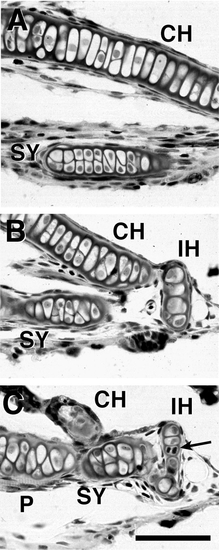
Chondrocyte arrangements in advanced larval cartilages. Horizontal 5-μm Epon sections through the pharynx at day 8, stained with methylene blue and basic fuchsin. The sections were selected from a serial series running ventral (A) to dorsal (C). Anterior is to left and medial is to the top. Chondrocyte cytoplasm is pale, such that the dark cell appositions and nuclei are prominent. Note the thin perichondrial layer around each of the cartilages and the chondrocyte in late telophase (arrow in C). Scale bar: 50 μm. |
Expression Data
Expression Detail
Antibody Labeling
Phenotype Data
Phenotype Detail
Acknowledgments
This image is the copyrighted work of the attributed author or publisher, and
ZFIN has permission only to display this image to its users.
Additional permissions should be obtained from the applicable author or publisher of the image.
Reprinted from Developmental Biology, 203, Kimmel, C.B., Miller, C.T., Kruse, G., Ullmann, B., BreMiller, R.A., Larison, K.D., and Snyder, H.C., The shaping of the pharnygeal cartilages during early development of the zebrafish, 245-263, Copyright (1998) with permission from Elsevier. Full text @ Dev. Biol.