
Fig. 5
- ID
- ZDB-FIG-090224-43
- Publication
- Yang et al., 2009 - hnRNP I Inhibits Notch Signaling and Regulates Intestinal Epithelial Homeostasis in the Zebrafish
- Other Figures
- All Figure Page
- Back to All Figure Page
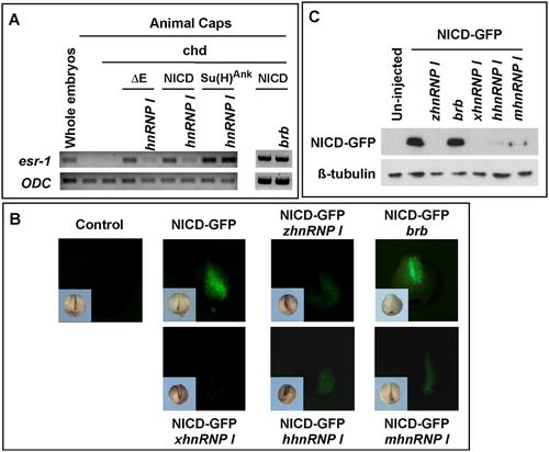
hnRNP I inhibits Notch signaling. (A) RT-PCR results demonstrate that hnRNP I (1 ng), but not brb (1 ng), inhibits the expression of esr-1 induced by Notch”E, NICD, but not that induced by Su(H)Ank in Xenopus animal caps. Animal caps were neuralized by chordin (Chd, 50 pg). (B) Stereoimages show hnRNP Is from the wild-type zebrafish (zhnRNP I, 2.5 ng), human (hhnRNP I, 2.5 ng), mouse (mhnRNP I, 2.5 ng) and Xenopus (xhnRNP I, 2.5 ng) decreased the level of NICD-GFP as revealed by the presence of green fluorescence. In contrast, the mutated form of hnRNP I in brom bones (brb) failed to reduce the level of NICD-GFP. Note that only one of the dorsal animal blastomeres, which later gave rise to the neural tissue, was injected. Inserts at the lower left corner of each panel are bright field images. (C) Western blot result showing the level of NICD-GFP (upper panel) was reduced by overexpression of hnRNP Is. Protein extracts were made from 15 injected embryos (20 ml lysis buffer per embryo). Each lane contains 10 ml lysate. After Western blot with the anti-GFP antibody, the membrane was re-probed with anti-tubulin antibody (lower panel). |