
Fig. 7
- ID
- ZDB-FIG-081111-36
- Publication
- Mendonsa et al., 1999 - Genetic analysis of tissue interactions required for otic placode induction in the zebrafish
- Other Figures
- All Figure Page
- Back to All Figure Page
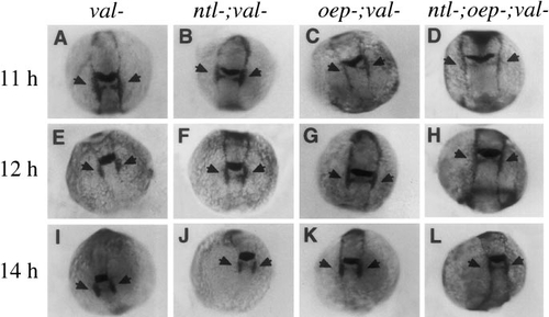
Disruption of hindbrain development in val- embryos does not further delay expression of dlx-3. Shown are expression patterns of both dlx-3 and krox-20 at (A–D) 11 h, (E–H) 12 h, and (I–L) 14 h. Although embryos lacking val function cannot be morphologically distinguished at the early developmental stages examined here, val- embryos were identified unambiguously by virtue of their low levels of krox-20 expression in rhombomere 5. Expression of dlx-3 is normal in val- (A, E, I) and ntl-;val- embryos (B, F, J). Upregulation of dlx-3 in the otic regions (arrows) in oep-;val- (C, G, K) and ntl-;oep-;val- embryos (D, H, L) is delayed to roughly the same extent as in oep- and ntl-;oep- embryos with normal val function (see Fig. 3). |
Reprinted from Developmental Biology, 206, Mendonsa, E.S. and Riley, B.B., Genetic analysis of tissue interactions required for otic placode induction in the zebrafish, 100-112, Copyright (1999) with permission from Elsevier. Full text @ Dev. Biol.