
Fig. 8
- ID
- ZDB-FIG-080925-30
- Publication
- Lee et al., 1999 - A wave of free cytosolic calcium traverses zebrafish eggs on activation
- Other Figures
- All Figure Page
- Back to All Figure Page
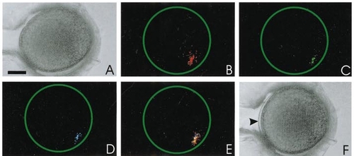
A representative example of a zebrafish egg with fluorescent microspheres loaded into its periphery. A and F are bright-field images taken before and after activation, respectively. A shows no separation between the chorion and the egg plasma membrane, whereas in F, the chorion is clearly raised (see arrowhead). B to D indicate confocal images taken at an optical plane containing the fluorescent microspheres. The fluorescent image in B was taken before the addition of 0.5% fructose in egg-water and then C and D at 2.5 and 5 min later, respectively. E shows the fluorescent images from B, C, and D superimposed on one another. This demonstrates that the egg does not rotate within its expanding chorion during the activation process. Scale bar is 200 μm. |
Reprinted from Developmental Biology, 214(1), Lee, K.W., Webb, S.E., and Miller, A.L., A wave of free cytosolic calcium traverses zebrafish eggs on activation, 168-180, Copyright (1999) with permission from Elsevier. Full text @ Dev. Biol.