FIGURE

Fig. S5

ID
ZDB-FIG-080902-9
Publication
König et al., 2007 - Splicing Segregation: The Minor Spliceosome Acts outside the Nucleus and Controls Cell Proliferation
Other Figures
All Figure Page
Back to All Figure Page
Fig. S5

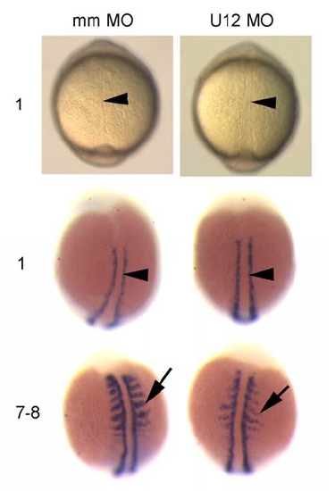
Developmental Patterning of the Midline and Somite Segmentation Are Unaffected in U12-MO-Injected Embryos during Early Somitogenesis
Top: Bright field view from dorsal onto 1 somite stage embryos. Arrowhead indicates the notochord. Middle: myoD expression in one somite stage embryos shown from dorsal view. Arrowhead indicates the notochord, purple staining indicates the paraxial mesoderm. No difference is seen between mismatch- (mm) and U12-morpholino (MO) injected embryos in the formation of the notochord or in myoD expression during the start of somitogenesis. Bottom: myoD expression in 7-8 somite stage embryos shown from dorsal view. Arrow indicates somite boundaries, which are formed but with reduced myoD staining in U12 MO injected embryos. Numbers on the left indicate somite numbers of the embryos shown.

Expression Data
Gene:
Fish:
Knockdown Reagent:
Anatomical Terms:
Stage Range: 1-4 somites to 5-9 somites

Expression Detail
Antibody Labeling
Phenotype Data

Phenotype Detail
Acknowledgments
This image is the copyrighted work of the attributed author or publisher, and ZFIN has permission only to display this image to its users. Additional permissions should be obtained from the applicable author or publisher of the image.

Reprinted from Cell, 131(4), König, H., Matter, N., Bader, R., Thiele, W., and Müller, F., Splicing Segregation: The Minor Spliceosome Acts outside the Nucleus and Controls Cell Proliferation, 718-729, Copyright (2007) with permission from Elsevier. Full text @ Cell