|
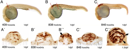
YFP expression in 1-dpf embryos of hoxa4a, hoxb4a, and hoxd4a enhancer detection lines as detected by immunohistochemistry. A-C: YFP expression pattern in the hindbrain and spinal cord of the transgenic lines CLGY409, CLGY838 and CLGY846. A′: Hoxa4a-YFP expression in elongated neuroepithelial cells in r8. The level of section is indicated in A. B′, B″: Transverse sections through the hindbrain of CLGY 838 show hoxb4a-YFP expression in elongated neuronal precursor cells on the level of r7 (B′) and r8, which also shows a neuron with a ventromedial projection (B″, black arrow). The levels of sections are indicated in B. C′: Many neuroepithelial cells were YFP positive in r7 of the hoxd4a enhancer detection embryos. The level of section is indicated in C. C″: Section through r7 of hoxd4a embryos at 1.5 dpf, showing YFP-positive postmitotic neurons, with young immature neurons sending out small primary projections (black arrows).
|