
FIGURE
Fig. S3
- ID
- ZDB-FIG-080828-56
- Publication
- Oteíza et al., 2008 - Origin and shaping of the laterality organ in zebrafish
- Other Figures
- All Figure Page
- Back to All Figure Page
Fig. S3
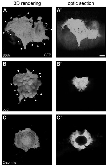
Mesenchymal-to-epithelial transition of the dorsal forerunner cell cluster. Three-dimensional renderings (A-C) and single focal planes through the centre of the DFC cluster (A′-C′) in a Tg(Sox17:GFP) embryo. At 80% epiboly (A,A′), migratory DFCs show numerous cell protrusions without clear directionality (arrowheads in A). At bud stage, cell protrusions start diminishing as the cluster rounds up (arrowheads in B) and organises into a more compact structure (B′). At the 2-somite stage, the DFC cluster has transformed into a vesicle (C) with a lumen at its centre (C′). Scale bar: 20 μm. |
Expression Data
| Gene: | |
|---|---|
| Fish: | |
| Anatomical Terms: | |
| Stage Range: | 75%-epiboly to 1-4 somites |
Expression Detail
Antibody Labeling
Phenotype Data
Phenotype Detail
Acknowledgments
This image is the copyrighted work of the attributed author or publisher, and
ZFIN has permission only to display this image to its users.
Additional permissions should be obtained from the applicable author or publisher of the image.
Full text @ Development