
FIGURE
Fig. 7
Fig. 7
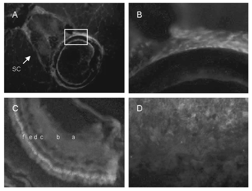
Cryosections of adult zebrafish labeled with VDR antibody visualized with Cy5 (red). Nuclei were labeled with DAPI (blue). A. Vertebra showing labeling in spinal cord (sc), vertebral body (boxed area), and surrounding muscle. B. Higher magnification of the vertebral body (boxed region). C, retina showing ganglion cell layer (a), inner plexiform layer (b), inner nuclear layer (c), outer plexiform layer (d), outer nuclear layer (e), and photo receptor layer (f). D, gills. |
Expression Data
| Antibody: | |
|---|---|
| Fish: | |
| Anatomical Terms: | |
| Stage: | Adult |
Expression Detail
Antibody Labeling
Phenotype Data
Phenotype Detail
Acknowledgments
This image is the copyrighted work of the attributed author or publisher, and
ZFIN has permission only to display this image to its users.
Additional permissions should be obtained from the applicable author or publisher of the image.
Reproduced from J Bone Miner Res 2008;23:1486-1496 with permission of the American Society for Bone and Mineral Research
Full text @ J. Bone Miner. Res.