
Fig. 2
- ID
- ZDB-FIG-080710-17
- Publication
- Wilson et al., 1991 - A pioneering growth cone in the embryonic zebrafish brain
- Other Figures
- All Figure Page
- Back to All Figure Page
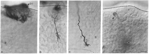
Micrographs from whole-mounted embryonic brains in which diI had been applied to the epiphysis (a-c) or to the DVDT (d). Rostral is to the right and dorsal is up. Dashed lines show the dorsal surface of the brain. (a) At 18 hr, cell bodies are labeled but none possesses axons or growth cones. (b) At 19.5 hr, a single pioneering growth cone is labeled approximately halfway down the DVDT. (c) At 21 hr, the pioneering growth cone is almost at the intersection with the TPOC (dotted line). (d) At 20 hr, a single neuron in the caudal epiphysis (arrowhead) is retrogradely labeled after diI application to the DVDT. E, epiphysis; DiI, application site. (Bar = 10 μm.) |