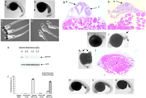
MO knockdown of gelsolin ventralizes embryos. Embryos 30 hpf after microinjection of control MO (2.5 ng/E; a) and gelsolin MO (b) are shown. Arrows indicate the location of the eyes. Embryos injected with control (c) or gelsolin MO (1 ng/E; d) are shown 72 hpf. Gelsolin MO resulted in ventralized embryos (b) and hatchlings with smaller and less pigmented eyes (d). The effect of different doses of gelsolin MO on the gelsolin and actin levels was determined by immunoblotting of embryo extracts prepared 8 h after MO injection (e; ref. 14). A representative immunoblot of four separate experiments is shown. The phenotypic effect of different doses of the gelsolin MO is summarized (f). Histological analyses using hematoxylin/eosin staining compare the normal and reduced development of anterior cephalic structures in control (g) and gelsolin MO-injected (h) embryos at 30 hpf. Locations of brain (B), eye (E), and yolk mass (Y) are indicated. Gelsolin mRNA (50 pg/E) injection results in dorsalized embryos (i and j). Arrows indicate duplicated posterior axial regions. Microinjection of human plasma gelsolin protein (4 ng/E) resulted in axis duplication (k). Arrows indicate duplicated anterior axial region including head and eyes. Histological section (l) of the embryo with duplicated axes showing notochord (nc) and optic cup (oc). Coinjection of gelsolin MO (2.5 ng/E) with 4 ng of human gelsolin protein (m), or with gelsolin mRNA (50 pg/E; n), or with chordin mRNA (50 pg/E; o) rescued the gelsolin MO-injected phenotypes. Embryos at 30 hpf are shown.
|
