
Fig. 10
- ID
- ZDB-FIG-080513-19
- Publication
- Herbomel et al., 2001 - Zebrafish early macrophages colonize cephalic mesenchyme and developing brain, retina, and epidermis through a M-CSF receptor-dependent invasive process
- Other Figures
- All Figure Page
- Back to All Figure Page
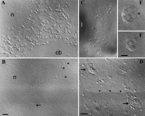
Apoptotic bodies accumulate in panther optic tectum and retina until eventual microglial colonization. Live embryos, 120 hpf, DIC optics. (A, B) Optic tectum, dorsal view, rostral upward, (A) Panther embryo. (B) Wild-type embryo; yellow shadows are due to xanthophores present above the brain (absent in panther); (arrow) blood vessel; (arrowheads) mitotic cells. (C) Retina. (D) Newly arrived early microglial cells (arrows) eliminating apoptotic bodies in the optic tectum; (arrowheads) midline. (E, F) Close-up on a tectal phagocyte at two depth planes, showing 7 still recognizable apoptotic bodies within its phagocytosed material; (asterisk) macrophage nucleus. (cb) cerebellum, (n) tectal neuropile. Bars: (A–C) 10 μm; (D) 10 μm; (E, F) 5 μm. |
Reprinted from Developmental Biology, 238(2), Herbomel, P., Thisse, B., and Thisse, C., Zebrafish early macrophages colonize cephalic mesenchyme and developing brain, retina, and epidermis through a M-CSF receptor-dependent invasive process, 274-288, Copyright (2001) with permission from Elsevier. Full text @ Dev. Biol.