FIGURE
Fig. 3
- ID
- ZDB-FIG-080424-48
- Publication
- Thisse et al., 2003 - Spatial and temporal expression patterns of selenoprotein genes during embryogenesis in zebrafish
- Other Figures
- All Figure Page
- Back to All Figure Page
Fig. 3
|
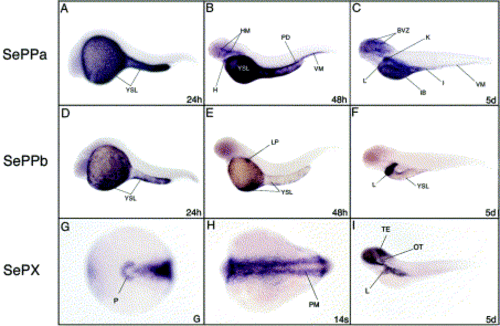
Expression patterns of the selenoprotein Pa (A–C), selenoprotein Pb (D–F) and methionine R-sulfoxide reductase (G–I) during development in zebrafish embryos. Development stage is indicated in the right lower corner (G, gastrula; 14s, 14 somites or in hours (h) or days (d) post-fertilization). BVZ, brain ventricular zone; H, heart; HM, head mesenchyme; I, intestine; IB, intestinal bulb; K, kidney; L, liver; LP, liver primordium; OT, otic vesicle; P, polster; PD, pronephric duct; PM, paraxial mesoderm; TE, tectum; VM, ventral mesenchyme; YSL, yolk syncytial layer. |
Expression Data
Expression Detail
Antibody Labeling
Phenotype Data
Phenotype Detail
Acknowledgments
This image is the copyrighted work of the attributed author or publisher, and
ZFIN has permission only to display this image to its users.
Additional permissions should be obtained from the applicable author or publisher of the image.
Reprinted from Gene expression patterns : GEP, 3(4), Thisse, C., Degrave, A., Kryukov, G.V., Gladyshev, V.N., Obrecht-Pflumio, S., Krol, A., Thisse, B., and Lescure, A., Spatial and temporal expression patterns of selenoprotein genes during embryogenesis in zebrafish, 525-532, Copyright (2003) with permission from Elsevier. Full text @ Gene Expr. Patterns

