
Fig. 7
- ID
- ZDB-FIG-080424-14
- Publication
- Westfall et al., 2003 - Requirement for intracellular calcium modulation in zebrafish dorsal-ventral patterning
- Other Figures
- All Figure Page
- Back to All Figure Page
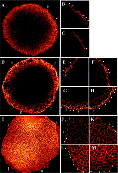
PI cycle inhibition increases the extent of nuclei containing β-catenin at sphere stage. Immunolocalization of β-catenin protein in sphere-stage embryos. Four-micron optical sections were collected with confocal microscopy. Embryos were oriented with the animal pole up. (A, D, and I) 20x magnification; (B, C, E–H, J–M) 63x magnification. The approximate locations of the higher magnification images are mapped, in lower case, onto the 20x image. (A) Wild-type embryo at sphere stage, 20x representation for orientation. Representative 63x images of the same embryo collected around the circumference are shown in (B) and (C) with dots noting the location of nuclear β-catenin. β-Catenin-positive nuclei are localized to a region of approximately one-third the circumference of the embryo. (D) XeC-injected embryo at 20x for orientation with individual high magnification images are shown in (E–H), dots noting intense nuclear β-catenin localization. XeC-treated embryos have expanded nuclear β-catenin domains that span more than 50% the circumference of the embryo. The L-690,330-injected embryo in (I) has nuclear β-catenin in ectopic locations that span the circumference of the embryo. Clusters of β-catenin-positive nuclei are present in (L) and (M) (L, M) β-Catenin localization at region opposite the location of (J) and (K). Numerous internal cells with nuclear β-catenin are not marked with dots. |
Reprinted from Developmental Biology, 259(2), Westfall, T.A., Hjertos, B., and Slusarski, D.C., Requirement for intracellular calcium modulation in zebrafish dorsal-ventral patterning, 380-391, Copyright (2003) with permission from Elsevier. Full text @ Dev. Biol.