
Fig. 2
- ID
- ZDB-FIG-080423-1
- Publication
- Lekven et al., 2003 - Wnt1 and wnt10b function redundantly at the zebrafish midbrain-hindbrain boundary
- Other Figures
- All Figure Page
- Back to All Figure Page
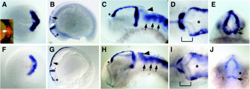
Comparison of wnt1 and wnt10b expression patterns. In situ hybridizations to detect wnt1 (A–E) or wnt10b (F–J) transcripts. In all images except (E) and (J), anterior is to the left. (A, F) Expression at 100% epiboly, dorsal view. Expression of both wnt1 and wnt10b forms a chevron pattern marking the prospective MHB. Inset in (A), double in situ for wnt8b (blue) and wnt1 (red). Arrow indicates region of nonoverlap at the midline. (B, G) Lateral views of embryos at the 16- somite stage. Expression of both genes marks the MHB and dorsal hindbrain/spinal cord (arrows), and both have anterior limits at the prospective epiphysis (asterisks). wnt10b expression is visible in the prospective cerebellum, but wnt1 is not (white arrows). (C–E, H–J) Embryos at approximately 30 hpf. (C, H) Lateral views of heads. wnt1 and wnt10b are both expressed from the epiphysis (asterisks) to the MHB. wnt10b is more strongly expressed at the posterior edge of the cerebellum (arrowheads), but both genes show enrichment in the rhombomeres (arrows). (D, I) Dorsal views of the same embryos as in (C, H). Brackets denote the MHB, and asterisks indicate the hindbrain ventricle. Note that wnt10b expression in the MHB appears slightly weaker than wnt1, but both are expressed on the anterior medial edge of the fold. (E, J) Posterior views of the MHBs of the same embryos as in (C, H). wnt10b expression appears generally weaker, but clear differences are seen in the ventral MHB where both genes are expressed in a stripe above the ventral midline (arrows), but wnt10b appears to be absent immediately above (asterisks). |
Reprinted from Developmental Biology, 254(2), Lekven, A.C., Buckles, G.R., Kostakis, N., and Moon, R.T., Wnt1 and wnt10b function redundantly at the zebrafish midbrain-hindbrain boundary, 172-187, Copyright (2003) with permission from Elsevier. Full text @ Dev. Biol.