Fig. 4
- ID
- ZDB-FIG-080422-25
- Publication
- Kurita et al., 2003 - Suppression of lens growth by alphaA-crystallin promoter-driven expression of diphtheria toxin results in disruption of retinal cell organization in zebrafish
- Other Figures
- All Figure Page
- Back to All Figure Page
|
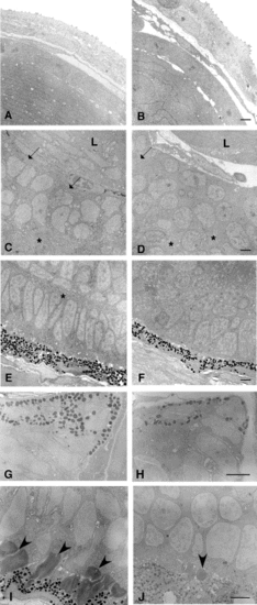
Electron microscopic analysis of the eye of zαAcry-DTA-expressing embryo. Electron micrographs of eyes of control (A, C, E, G, I) and zαAcry-DTA-injected (B, D, F, H, J) zebrafish embryos at 54 hpf (A–F) and 72 hpf (G–J). (A, B) Cornea and lens at 54 hpf. Morphology of the cornea and lens epithelial cells is essentially the same between control (A) and zαAcry-DTA-injected (B) embryos. Many thin lens fiber cells are seen in the control eye (A), and thick cells with a large round nucleus are seen in the zαAcry-DTA-injected (B) embryos. (C, D) Interface between the lens (L) and the retina. Bundles of many cell processes (arrows) are seen in the innermost layer of the control retina at 54 hpf. At the bottom of the photograph (* in C), a continuous layer of cell processes is seen. In the retina of the zαAcry-DTA-injected animals (D), cell processes do not form continuous layers and some are seen within small areas (* in D) between cell nuclei. (E, F) Outermost area of the retina. A layer of regularly arranged columnar nuclei is separated by a layer of cell processes (*) from the inner cell nuclei in the control (E). No regular arrangement of the nuclei can be seen the eye of the zαAcry-DTA-injected embryos (F). Retinal pigment epithelial cells of both the zαAcry-DTA-injected (F) and the control (E) embryo contain pigment granules. (G, H) Tip of the eye cup at 72 hpf. Pigmented cells at the tip of the eye cup expand toward the lens in both control (G) and the zαAcry-DTA-injected (H) embryos. (I, J) Outermost area of the retina at 72 hpf. Photoreceptor outer segments are observed in the control embryos (I). In contrast, outer segment-like structures were seen only occasionally in the zαAcry-DTA-injected embryos (J). Outer segments (I) or outer segment-like structure (J) are indicated by arrowheads. Scale, 2 μM. |
Reprinted from Developmental Biology, 255(1), Kurita, R., Sagara, H., Aoki, Y., Link, B.A., Arai, K.-I., and Watanabe, S., Suppression of lens growth by alphaA-crystallin promoter-driven expression of diphtheria toxin results in disruption of retinal cell organization in zebrafish, 113-127, Copyright (2003) with permission from Elsevier. Full text @ Dev. Biol.

