
Fig. 1
- ID
- ZDB-FIG-080417-44
- Publication
- Fadool, 2003 - Development of a rod photoreceptor mosaic revealed in transgenic zebrafish
- Other Figures
- All Figure Page
- Back to All Figure Page
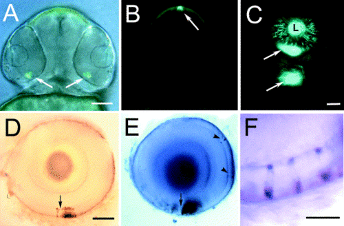
Comparison of EGFP and rhodopsin expression in transgenic zebrafish. EGFP expression in photoreceptor cells (arrows) of the ventral retina (A) and the pineal (B) of transgenic zebrafish larva observed at 60 hpf. (C) At 84 hpf, EGFP fluorescence could be observed in the ventral patch (arrows) of the retinas and in cells sporadically positioned across the retina. Apparent fluorescence by the lens (L) is due to photoreceptor cell expression. In situ hybridization of EGFP (D) and rhodopsin (E) demonstrate a similar pattern of labeling of cells in the outer nuclear layer (ONL) of the ventral retina and on opposite sides of the choroid fissure (arrow). Sporadic labeling of cells in the ONL is also observed with the opsin probe (arrowheads). (F) Double labeling demonstrated colocalization of the EGFP transcript (brown) and rhodopsin transcript (purple) to sporadically spaced cells located in the outer nuclear layer of larval fish. Bar, 50 mm in (A–C); 25 mm in (D, E); 10 mm in (E). |
Reprinted from Developmental Biology, 258(2), Fadool, J.M., Development of a rod photoreceptor mosaic revealed in transgenic zebrafish, 277-290, Copyright (2003) with permission from Elsevier. Full text @ Dev. Biol.