
Fig. 4
- ID
- ZDB-FIG-080402-27
- Publication
- De Mazière et al., 2008 - Egfl7 knockdown causes defects in the extension and junctional arrangements of endothelial cells during zebrafish vasculogenesis
- Other Figures
- All Figure Page
- Back to All Figure Page
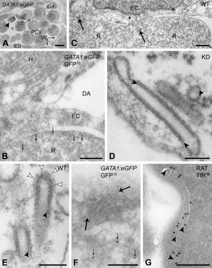
Characteristic striated tubules are exclusively found in red blood cell precursors. A,B,F,G: Ultrathin cryosections. C-E: Epon sections. A: Axial vascular area of 24 hours postfertilization (hpf) GATA1:eGFP transgenic embryo, labeled for green fluorescent protein (GFP). Arrowhead, apoptotic cell. B: Higher magnification of area boxed in A. Red blood cell precursor located between dorsal aorta (DA) and posterior cardinal vein (PCV), and identified by labeling with anti-GFP antibodies (10-nm gold particles, GFP10, small arrows). C-E: Characteristic tubules (arrows) near the plasma membrane of red blood cell precursors in control (C,E) and Egfl7 knockdown (D) embryos display regular cross-striations and an inner coating (arrowheads) of the limiting membrane. Open arrowheads, clathrin coat. F: Striated tubules (arrows) with electron-dense inner coating inside the electron-lucent limiting membrane colocalize with GFP label (10-nm gold particles, GFP10, small arrows) in red blood cell precursors of GATA1:eEGFP transgenic embryos. G: Rat bone marrow erythroblast. Intracellular tubules with inner coating (arrowheads), resembling zebrafish striated tubules, are labeled with 10-nm gold particles for transferrin receptor (TfR10). EC, endothelial cell; ED, endoderm; H, hypochord; R, red blood cell precursor; VE, venous endothelium. Scale bars = 5 μm in A, 0.5 μm in B, 1 μm in C, 100 nm in D,E, 200 nm in F,G. |