
Fig. 5
- ID
- ZDB-FIG-080326-94
- Publication
- Sato-Maeda et al., 2008 - Position fine-tuning of caudal primary motoneurons in the zebrafish spinal cord
- Other Figures
- All Figure Page
- Back to All Figure Page
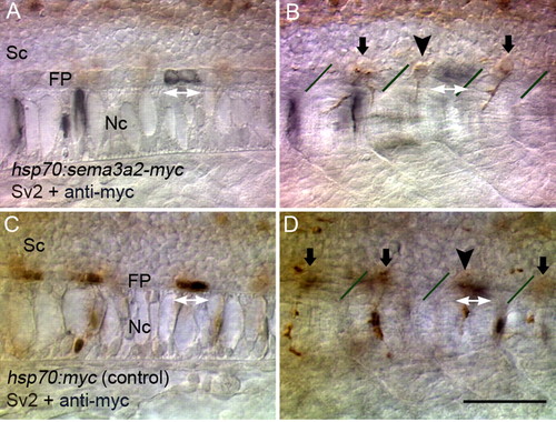
Abnormal CaP cell positioning induced by focal ectopic Sema3ab. (A,B) Optical sections at different focal levels of an embryo in which Sema3ab-Myc was transiently expressed. Ectopic Sema3ab was labeled with anti-Myc (black) and CaPs with mAb Sv2 (brown). In a case in which ectopic Sema3ab was expressed in floor plate cells (white double arrows in A), the CaP cell body was shifted anteriorly (arrowhead in B) away from the cells expressing Sema3ab. (C,D) Optical sections of a control embryo. Myc epitope expression (white double arrows in C) did not affect the location of the CaP cell body (arrowhead in D) and overlapped with it. Lines in B and D indicate somitic borders. Sc, spinal cord; FP, floor plate; Nc, notochord. Scale bar: 50 μm. |