
Fig. 3
- ID
- ZDB-FIG-080326-107
- Publication
- Devine et al., 2008 - Robo-Slit interactions regulate longitudinal axon pathfinding in the embryonic vertebrate brain
- Other Figures
- All Figure Page
- Back to All Figure Page
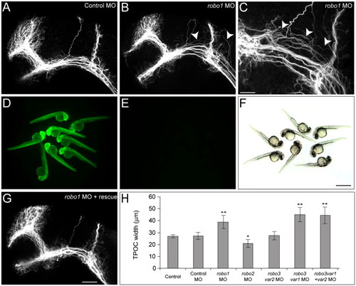
Aberrant dorsal spreading of the TPOC is observed following knockdown of Robo1. Dissected brains of 28 hpf zebrafish embryos labeled with acetylated α-tubulin to show all axons. Rostral is to the left and dorsal is to the top. (A) In control embryos the scaffold of axon tracts develops normally. Following injection of 5 ng robo1 MO2, TPOC axons were observed to spread inappropriately into more dorsal regions of the mesencephalon (B and C; arrowheads). (D–G) The specificity of the robo1 MO knockdown was verified by loss of GFP fluorescence following coinjection of 250 pg 5′ robo1-GFP cRNA and 5 ng robo1 MO2 (E, bright field in panel F) and rescue of the TPOC phenotype by coinjection with 250 pg of robo1-rescue cRNA (G). (H) The severity of the TPOC spreading phenotype was quantified by measuring the width of the TPOC. Values represent the mean ± SD. Average TPOC width measurements were analyzed using ANOVA, followed by the Tukey HSD test, *P < 0.05, **P < 0.001. Scale bars: A, B, G: 50 μm; C: 20 μm; D–F: 400 μm. |
| Fish: | |
|---|---|
| Knockdown Reagent: | |
| Observed In: | |
| Stage: | Prim-5 |
Reprinted from Developmental Biology, 313(1), Devine, C.A., and Key, B., Robo-Slit interactions regulate longitudinal axon pathfinding in the embryonic vertebrate brain, 371-383, Copyright (2008) with permission from Elsevier. Full text @ Dev. Biol.