
Fig. S3
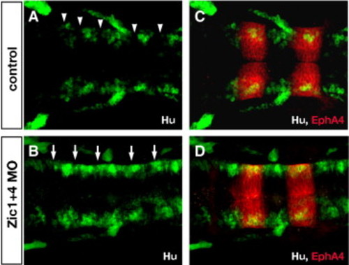
Zic1 and Zic4-deficient embryos lack normal segmental patterns of neuronal differentiation in the hindbrain. (A,B) Longitudinal confocal sections through the dorsal hindbrain of Hu immunolabeling (green, labels post-mitotic neurons), and (C,D) of double labeling with antibodies to Hu (green) and EphA4 (red, labels r3 and r5). In 24 hpf control embryos (A,C) post-mitotic neurons are clustered in the center of rhombomeres and absent from rhombomere boundaries (white arrowheads in A), while in Zic1 + 4 morphants (B,D) segmental pattern of Hu labeling is disrupted with ectopic differentiation of Hu-positive neurons in boundary regions (white arrows in B). |
| Fish: | |
|---|---|
| Knockdown Reagents: | |
| Observed In: | |
| Stage: | Prim-5 |
Reprinted from Developmental Biology, 314(2), Elsen, G.E., Choi, L.Y., Millen, K.J., Grinblat, Y., and Prince, V.E., Zic1 and Zic4 regulate zebrafish roof plate specification and hindbrain ventricle morphogenesis, 376-392, Copyright (2008) with permission from Elsevier. Full text @ Dev. Biol.