
Fig. S6
- ID
- ZDB-FIG-080211-29
- Publication
- Filippi et al., 2007 - Expression and function of nr4a2, lmx1b, and pitx3 in zebrafish dopaminergic and noradrenergic neuronal development
- Other Figures
- All Figure Page
- Back to All Figure Page
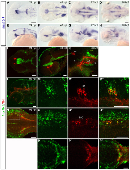
lmx1b.1 is co-expressed with TH in the NA neurons of the locus coeruleus and medulla oblongata. (A-H) Whole mount in situ hybridization showing lmx1b.1 expression pattern at 24 hpf (A, E), 48 hpf (B, F), 72 hpf (C, G) and 96 hpf (D, H). Dorsal (A-D) and lateral (E-H) views of the head are represented, anterior is to the left. (I-P″) Confocal z-projections of whole mount FISH to lmx1b.1 (green) combined with anti-TH immunohistochemistry (red) representing the spatial relationship between lmx1b.1-expressing and THir cells. (I) Dorsal view (35 μm projection) of the head at 24 hpf. (J) Lateral view (17 μm projection) of a 48 hpf embryo. (K) High magnification of the diencephalic area (40 μm projection) in a 96 hpf embryo (approximate area framed in H). No co-expression of lmx1b.1 and TH is detected in the DA neurons of the posterior tuberculum. Double labelling is instead observed in the NA neurons of the locus coeruleus (L-M″) and medulla oblongata (N-O"), as well as of the area postrema (P-P″). (L) Single confocal image at the level of the locus coeruleus of a 96 hpf embryo. High magnification of the framed area is showed in M-M″ (23 μm projection). (N) Dorsal overview of the medulla oblongata in a 96 hpf embryo (4 μm projection). High magnification of the framed area is showed in O-O″ (single plane). (P-P″) 3 μm dorsal projection through the DA neurons of the area postrema. I, N, O-O″ dorsal views; J, K, L, M-M″ lateral views; anterior is to the left. Scale bars in A is for A-C, E-G and in D is for D, H: 100 μm. Scale bars in I-P″: 50 μm. |