
FIGURE
Fig. 3
Fig. 3
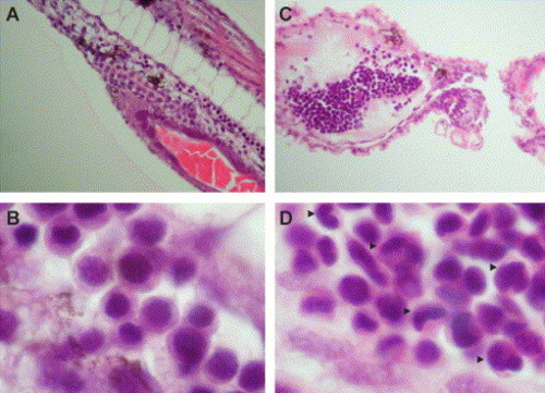
H&E sectioning of WT (A, B) and ChdMO embryos (C, D) at low (A, C) and high power (B, D) magnification at 48 hpf. In WT embryos, there was no defined population of cells in the ICM although erythroid cells can be readily identified at the axial circulation (B). In the chordin morphants, the ICM remained expanded but instead of a monotonous population seen at 24 hpf, the cells in ICM at 48 hpf became heterogeneous. Monocyte-looking cells with horseshoe-shaped nuclei are readily encountered (arrowheads). |
Expression Data
Expression Detail
Antibody Labeling
Phenotype Data
Phenotype Detail
Acknowledgments
This image is the copyrighted work of the attributed author or publisher, and
ZFIN has permission only to display this image to its users.
Additional permissions should be obtained from the applicable author or publisher of the image.
Reprinted from Developmental Biology, 277(1), Leung, A.Y., Mendenhall, E.M., Kwan, T.T., Liang, R., Eckfeldt, C., Chen, E., Hammerschmidt, M., Grindley, S., Ekker, S.C., and Verfaillie, C.M., Characterization of expanded intermediate cell mass in zebrafish chordin morphant embryos, 235-254, Copyright (2005) with permission from Elsevier. Full text @ Dev. Biol.