
Fig. 7
- ID
- ZDB-FIG-071004-21
- Publication
- Uemura et al., 2005 - Comparative functional genomics revealed conservation and diversification of three enhancers of the isl1 gene for motor and sensory neuron-specific expression
- Other Figures
- All Figure Page
- Back to All Figure Page
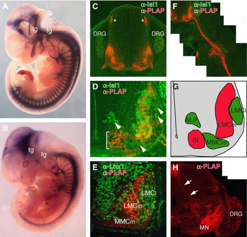
Functional conservation and diversification of CREST2 for motor and sensory neuron-specific gene expression in mouse. (A and B) Lateral views of the E11.5 Tg(zCREST2/isl1-PLAP) embryo (A) and Tg(hCREST2/isl1-PLAP) embryo (B). In both embryos, PLAP was expressed in spinal motor neurons that extend their axons ventrally, while PLAP expression in the sensory neurons, such as trigeminal ganglion neurons (tg), facial ganglion neurons (fg), and dorsal root ganglion neurons (DRG), was observed only in the Tg(zCREST2/isl1-PLAP) embryo (see also C and H). (C, D, and F) Cross-sections at the brachial level of E11.5 Tg(zCREST2/isl1-PLAP) embryos doubly labeled with anti-Isl1 antibody (green) and anti-PLAP antibody (red) showing PLAP expression in the subset of spinal motor neurons innervating the ventral muscles of the limb and in DRG neurons. Asterisks in C indicate the dorsal root entry zone (DREZ) where the central axons of the DRG neurons enter the spinal cord. Close-up view of the ventral spinal cord (D) showing expression of PLAP in the specific subset of spinal motor neurons, excluding the dorsomedially, ventromedially, and dorsolaterally (arrowheads) located Isl1-positive motor neurons. In this particular line, ectopic PLAP expression was also observed in the ventralmost region of the spinal cord (bracket). The specific projection of PLAP-positive motor axons into the ventral limb bud is shown in F. (E) Double labeling with anti-Lhx1 antibody (green) and anti-PLAP antibody (red) at the brachial level of the E11.5 Tg(zCREST2/isl1-PLAP) embryo revealed that PLAP-positive motor neurons were negative for Lhx1, a marker for LMCl motor neurons, supporting their identity as LMCm motor neurons. Lhx1 was also expressed in other interneurons (Tsuchida et al., 1994). (G) Schematic illustration of D. Regions containing PLAP-positive and PLAP-negative neurons are indicated in red and green, respectively. M.P., motor neuron precursors; IN, interneurons. (H) Brachial level of the E14.5 Tg(zCREST2/isl1-PLAP) embryo labeled with anti-PLAP antibody (red). The PLAP-positive Ia afferent fibers (arrows) were observed penetrating the spinal cord and projecting ventrally to the motor neurons (MN) in the ventral horn. |
Reprinted from Developmental Biology, 278(2), Uemura, O., Okada, Y., Ando, H., Guedj, M., Higashijima, S., Shimazaki, T., Chino, N., Okano, H., and Okamoto, H., Comparative functional genomics revealed conservation and diversification of three enhancers of the isl1 gene for motor and sensory neuron-specific expression, 587-606, Copyright (2005) with permission from Elsevier. Full text @ Dev. Biol.