
Fig. 1
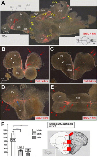
Proliferation sites in the zebrafish adult telencephalon. (A–E) BrdU immunocytochemistry (red staining) on brain sections of 6-month-old adult zebrafish sacrificed 4 h after 2 BrdU injections. Vibratome sections are observed under conventional fluorescence microscopy, (A) parasagittal section, anterior left, (B–D) cross-sections at increasingly posterior levels (indicated in panel A), dorsal up, (E) horizontal section (level indicated in panel A), anterior left. In panel A, intensely labeled sites are numbered (red arrows: telencephalon, yellow arrowheads: other domains). Note the stripe of intense staining within the ventral subpallium (numbered 1 in all sections). Other sites: (2) optic tracts, (3) ventricular zone of the hypothalamus, (4) anterior thalamus, (5) periventricular gray zone, (6) torus longitudinalis, (7) cerebellar external granular layer, (8) valvula cerebelli, (9) lobus caudalis cerebelli. Nonventricular proliferation is also visible in all cross-sections and indicated by white arrowheads. The only staining visible in the OB (E, red arrowheads) likely corresponds to glia in the superficial olfactory nerve layer (Byrd and Brunjes, 2001). In the posterior telencephalon, prominent proliferation is observed at the junction between the dorsal and ventral domains (blue arrow in panel D) and in the posterior pallium (red arrow numbered 1p in panel D). (F) Number of BrdU-positive cells in the telencephalon along the DV axis, counted in the areas boxed in red in the scheme on the right panel (Wullimann et al., 1996). Ventral subpallium: 1245 ± 245 BrdU-positive cells/mm3, dorsal subpallium: 312 ± 136/mm3, medial pallium: 138 ± 37/mm3 (on a total of 9 sections from 3 different fish at anterior telencephalic levels, all areas containing a comparable total number of cells, statistical analysis performed using independent samples Student′s t test). Abbreviations, after Wullimann et al. (1996): CCe: corpus cerebellaris, EGL: external granular layer, Hyp: hypothalamus, IGL: internal granular layer, LCa: lobus caudalis cerebelli, Pa: pallium, PGZ: periventricular gray zone, OB: olfactory bulb, OT: optic tract, Pn: pons, Sub: subpallium, Te: telencephalon, TeO: tectum opticum, TL: torus longitudinalis, V: posterior domain of the ventral telencephalon, val: valvula cerebelli. |
Reprinted from Developmental Biology, 295(1), Adolf, B., Chapouton, P., Lam, C.S., Topp, S., Tannhauser, B., Strähle, U., Gotz, M., and Bally-Cuif, L., Conserved and acquired features of adult neurogenesis in the zebrafish telencephalon, 278-293, Copyright (2006) with permission from Elsevier. Full text @ Dev. Biol.