
FIGURE
Fig. 5
- ID
- ZDB-FIG-070514-5
- Publication
- Li et al., 2007 - Cloning and spatial and temporal expression of the zebrafish dopamine D1 receptor
- Other Figures
- All Figure Page
- Back to All Figure Page
Fig. 5
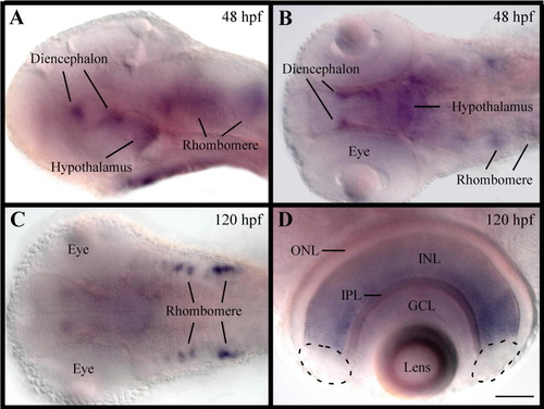
Expression of drd1 in developing embryos. A-C: Lateral and dorsal views of developing embryos at 48 hours postfertilization (hpf) and 120 hpf. Anterior is to the left. Note the increase of drd1 expression in the late developmental stages. D: Expression of drd1 in the retina at 120 hpf. Expression of drd1 was detected throughout the entire inner nuclear layer with the exception of the marginal zones (outlined by dashed lines). ONL, outer nuclear layer; INL, inner nuclear layer; IPL, inner plexiform layer; GCL, ganglion cell layer. Scale bar = 70 μm in A-C, 35 μm in D. |
Expression Data
| Gene: | |
|---|---|
| Fish: | |
| Anatomical Terms: | |
| Stage Range: | Long-pec to Day 5 |
Expression Detail
Antibody Labeling
Phenotype Data
Phenotype Detail
Acknowledgments
This image is the copyrighted work of the attributed author or publisher, and
ZFIN has permission only to display this image to its users.
Additional permissions should be obtained from the applicable author or publisher of the image.
Full text @ Dev. Dyn.