
Fig. 7
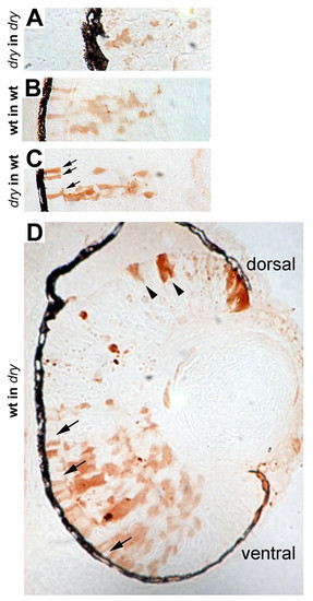
Genetic mosaic analysis of disarrayed retinal cells. Genetic mosaic retinas at 72 hpf where donor cells were lineage-labeled using biotin conjugated dextran (brown). (A) disarrayed mutant cells in a disarrayed mutant host embryo. Note reduced morphological differentiation and lamination of cells. (B) Wild-type cells in a wild-type host embryo. (C) disarrayed mutant cells in a wild-type host embryo. The disarrayed cells show wild-type morphology and lamination in the wild-type environment (compare B and C). For example, morphological rescue of photoreceptor elongation can be seen (C, arrows). (D) Wild-type cells in a disarrayed mutant embryo. Small wild-type cell clones resemble mutant cells as they appear delayed in morphological differentiation and lamination (dorsal, arrowheads). Wild-type cells in clones with greater donor mosaicism have normal morphological differentiation and rescue adjacent disarrayed mutant cells (ventral, arrows). The number of mosaic embryos examined: n = 2 disarrayed into disarrayed, n = 5 wild-type into wild-type, n = 9 disarrayed into wild-type, n = 8 wild-type into disarrayed. Each embryo contained multiple cell clones. |