
Fig. 1
- ID
- ZDB-FIG-070331-1
- Publication
- Rich et al., 2007 - Kit-like immunoreactivity in the zebrafish gastrointestinal tract reveals putative ICC
- Other Figures
- All Figure Page
- Back to All Figure Page
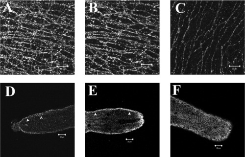
Anti-Kit rabbit polyclonal antibody identifies putative ICC networks in the adult zebrafish GI tract. Full thickness confocal stack reconstruction of an adult zebrafish GI whole mount preparation immunostained using rabbit anti-Kit polyclonal antibody (A). Total thickness of reconstructed stack ≈20 μm. Reconstruction of a partial thickness stack (≈8-μm-thick) shows a kit-positive network in the myenteric plexus region with stellate-shaped, highly branching cells (B). A second distinct network of slender bipolar cells oriented parallel to the circular smooth muscle cells was located closer to the submucosal border and is shown in a partial stack reconstruction of a 6-μm-thick region (C). Kit-like immunoreactivity was observed in the GI tract of zebrafish larvae beginning at 7 dpf. Single confocal sections approximately 0.6-μm-thick of mid-saggital sections from the posterior end of the larvae GI tract show branching cells with slender processes in the tunica muscularis (D, arrows). Background staining of the mucosal layer prevented full-thickness stack reconstruction. The density of Kit-positive cells was increased at 11 dpf (E), and a network of Kit-positive cells was observed at 20 dpf (F). Scale bars = 20 μm. |