|
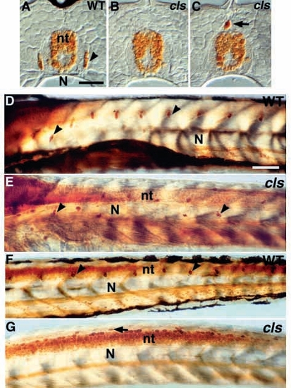
DRG sensory neurons are disrupted in cls- larvae. (A-C) Transverse section of dorsal tail of (A) wild-type larva at 7 dpf stained with anti-Hu antibody reveals small clusters of Hu-positive neurons (arrowhead) in DRG. These are usually absent from the tail of cls- homozygotes (B). cls- larvae have an increased frequency of extramedullary cells dorsal to the neural tube (nt; arrow in C and G). (D-G) Lateral views of whole-mount larvae at 3 dpf stained with anti-Hu antibody reveal a segmental pattern of DRGs (arrowheads) throughout the trunk (D) and tail (F), but these cells are reduced and misplaced in the trunk (E) and absent from the tail (G) of cls- larvae. N, notochord; sc, spinal cord. Scale bar, 25 μm (A-C) and 75 μm (D-G).
|