
Fig. 5
- ID
- ZDB-FIG-070219-8
- Publication
- Ernest et al., 2007 - Localization of anosmin-1a and anosmin-1b in the inner ear and neuromasts of zebrafish
- Other Figures
- All Figure Page
- Back to All Figure Page
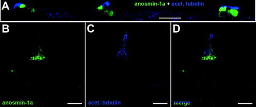
Expression of anosmin-1a in sensory hair cells of the neuromast. (A) Confocal sections of the three terminal neuromasts showing anosmin-1a staining (green) in the hair bundle and, at a lower level, in the body of sensory hair cells. Kinocilia were labeled with an anti-acetylated tubulin antibody (blue). (B–D) Higher magnification of a single neuromast revealed a strong anosmin-1a accumulation in the stereocilia of hair cells and, to a lesser extent, in the cell body and in the kinocilia (lower half) which was co-labeled using an anti-acetylated tubulin antibody. Data represent the maximum intensity projection of the z-sections of the acquired z-stacks (A) or maximum intensity projections of the acquired z-stacks (B–D). Scale bar: 20 μm (A), 10 μm (B–D). |
| Gene: | |
|---|---|
| Fish: | |
| Anatomical Term: | |
| Stage: | Day 4 |
Reprinted from Gene expression patterns : GEP, 7(3), Ernest, S., Guadagnini, S., Prevost, M.C., and Soussi-Yanicostas, N., Localization of anosmin-1a and anosmin-1b in the inner ear and neuromasts of zebrafish, 274-281, Copyright (2007) with permission from Elsevier. Full text @ Gene Expr. Patterns