
Fig. 5
- ID
- ZDB-FIG-061121-46
- Publication
- Chapouton et al., 2006 - her5 expression reveals a pool of neural stem cells in the adult zebrafish midbrain
- Other Figures
- All Figure Page
- Back to All Figure Page
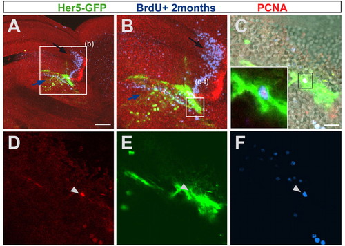
her5-positive cells are self-renewing. A 3-month-old her5:gfp transgenic brain was injected cumulatively over 9 days with BrdU and sacrificed 2 months later. The sagittal section is seen as an overview in A, and magnified in B-F, as single confocal planes. (C-F) Enlargement on an Her5-GFP cell (green, pointed by an arrowhead) that has incorporated BrdU 2 months earlier (blue) and is PCNA-positive (red), indicating that it has remained in a proliferative state after division, i.e. it is a self-renewing progenitor. Note that most cells of the IPZ that have incorporated BrdU 2 months earlier move towards ventral positions into the tegmentum (see in B the location of blue cells below the Her5-GFP domain, blue arrow), while cells originating from the tectal proliferation zone are displaced towards anterior into the optic tectum (B, black arrow). Scale bars: 100 μm in A; 10 μm in C. |