
FIGURE
Fig. 7
- ID
- ZDB-FIG-060628-6
- Publication
- Kudo et al., 2004 - Zebrafish periostin is required for the adhesion of muscle fiber bundles to the myoseptum and for the differentiation of muscle fibers
- Other Figures
- All Figure Page
- Back to All Figure Page
Fig. 7
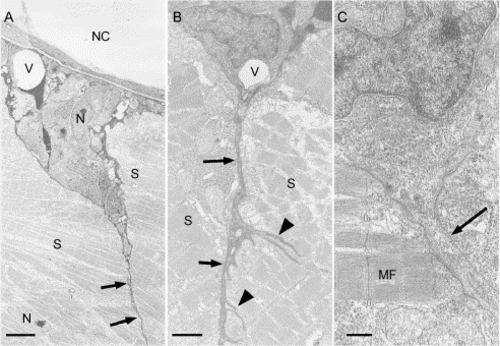
Structure of the transverse myosepta in wild-type embryos at 48 hpf. The triangular regions surrounded by adjacent myotomes and notochord or spinal cord. Myosepta are recognized as cytoplasmic processes (arrows in A and B), which are extended from cells in the triangular regions; and these cytoplasmic processes have small secondary protrusions (arrowheads in B). Cell membranes (an arrow in C) in the cytoplasmic process of myoseptum can be obviously discernible. Abbreviations: M, myotome; MF, myofibers; N, nucleus; NC, notochord; V, blood vessel. Scale bars: A, 6 μm; B, 1.5 μm; C, 0.7 μm. |
Expression Data
Expression Detail
Antibody Labeling
Phenotype Data
Phenotype Detail
Acknowledgments
This image is the copyrighted work of the attributed author or publisher, and
ZFIN has permission only to display this image to its users.
Additional permissions should be obtained from the applicable author or publisher of the image.
Reprinted from Developmental Biology, 267(2), Kudo, H., Amizuka, N., Araki, K., Inohaya, K., and Kudo A., Zebrafish periostin is required for the adhesion of muscle fiber bundles to the myoseptum and for the differentiation of muscle fibers, 473-487, Copyright (2004) with permission from Elsevier. Full text @ Dev. Biol.