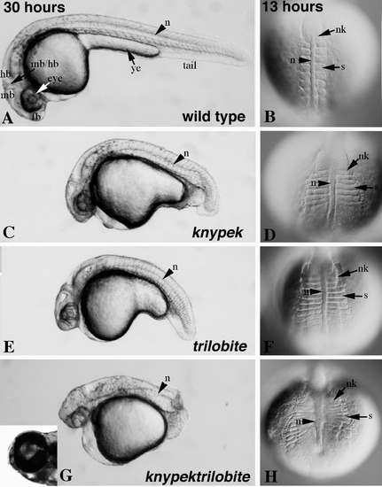
Fig. 2
- ID
- ZDB-FIG-060216-8
- Publication
- Marlow et al., 1998 - Functional interactions of genes mediating convergent extension, knypek and trilobite, during the partitioning of the eye primordium in zebrafish
- Other Figures
- All Figure Page
- Back to All Figure Page
|
knym119 trim209 double-mutant embryos exhibit additive defects in convergent extension during somitogenesis and 1 dpf, and a synergistic cyclopia defect, compared to single mutants. (A, C, E, G) Dissecting microscope images, lateral view of live embryos at 30 hpf. The length of the embryo is decreased in knym119 (C) and trim209 (E) mutants and dramatically decreased in the double mutant (G) compared to wild type (A). Abnormal shape of the yolk cell and cyclopia are seen in the double mutant (G, inset). (B, D, F, H) Dorsal views of live embryos at 13 hpf using Nomarski optics. The mediolateral width and AP extension of somites (s), notochord (n), and neural keel (nk) are affected in an additive fashion in double mutants (H) compared to knym119 (D) and trim209 (F) single-mutant embryos. fb, forebrain; hb, hindbrain; mb, midbrain; mb/hb, midbrain–hindrain boundary; ye, yolk extension. |
| Fish: | |
|---|---|
| Observed In: | |
| Stage Range: | 5-9 somites to Prim-15 |
Reprinted from Developmental Biology, 203, Marlow, F., Zwartkruis, F., Malicki, J., Neuhauss, S.C.F., Abbas, L., Weaver, M., Driever, W., and Solnica-Krezel, L., Functional interactions of genes mediating convergent extension, knypek and trilobite, during the partitioning of the eye primordium in zebrafish, 382-399, Copyright (1998) with permission from Elsevier. Full text @ Dev. Biol.

