
Fig. 4
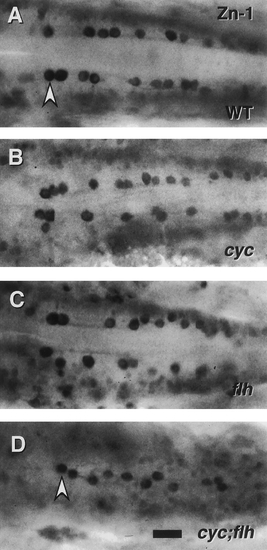
Patterning of ic interneurons is severely disrupted in cyc;flh double mutants. Dorsal views of the caudal hindbrain, with anterior to the left, in whole-mounted embryos labeled with the zn-1 monoclonal antibody at 24 hr. This antibody binds a neuronal antigen that is prominently expressed by ic interneurons, a distinctive class of neurons cells present in the caudal hindbrain that project axons ipsilaterally and caudally, within the medial longitudinal fascicles, to the spinal cord (Mendelson, 1986). In the wild type (A) the ic neurons (arrowhead) line up in bilateral rows adjacent to the medial longitudinal fascicles. The same two cell rows are present (and are closer together) in cyc (B) and flh (C) single mutants. Only a single disorganized row of ic cells is present, just at the midline, in the cyc;flh double mutant (D). Scale bar, 25 um. |
Reprinted from Developmental Biology, 187(2), Halpern, M.E., Hatta, K., Amacher, S.L., Talbot, W.S., Yan, Y.-L., Thisse, B., Thisse, C., Postlethwait, J.H., and Kimmel, C.B., Genetic interactions in zebrafish midline development, 154-170, Copyright (1997) with permission from Elsevier. Full text @ Dev. Biol.