
Fig. 5
- ID
- ZDB-FIG-051025-5
- Publication
- Flanagan-Steet et al., 2005 - Neuromuscular synapses can form in vivo by incorporation of initially aneural postsynaptic specializations
- Other Figures
- All Figure Page
- Back to All Figure Page
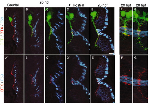
Distinct modes of synapse formation on muscle pioneers and fast fibers. Cross-sections through HB9:GFP fish at 20 hpf (A-D,F) or 28 hpf (E,G) were stained with anti-myosin (F59) and BTX. (A-D) CaP extends to contact muscle pioneers as the other adaxial fibers migrate radially. (E) CaP then extends past the pioneers as fast, weakly F59-immunoreactive, fibers form. AChR clusters on migrating adaxial cells are lost and new clusters form on fast fibers as, or after, they are contacted by CaP. As in Figs 1 and 2, the rostrocaudal sequence of development allows the visualization of many stages in a single fish. (F,G) En face views at 20 hpf and 28 hpf to show that AChR clusters on muscle pioneers are larger and more elongated than those on fast fibers. A'-G' show F59 and BTX, but not axons, to highlight AChR cluster position and morphology. Scale bar in E: 10 µm for all panels. |