
FIGURE
Fig. S2
- ID
- ZDB-FIG-050729-11
- Publication
- Xiao et al., 2005 - A GFP-based genetic screen reveals mutations that disrupt the architecture of the zebrafish retinotectal projection
- Other Figures
- All Figure Page
- Back to All Figure Page
Fig. S2
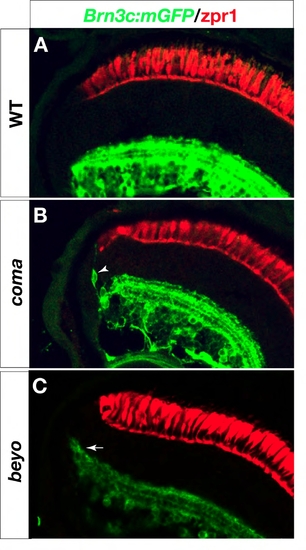
Retina defects in coming apart (coma) and beyond borders (beyo) mutants. Six dpf retina sections labeled with anti-GFP in green and zpr1 in red (A-C). (B) In coma, dendrite-bearing, GFP-positive neurons (arrowhead) are found at the margins of the inner nuclear layer. (C) In beyo, the edge of the inner plexiform layer ?smiles?, i. e. it bends toward the outer retina (arrow). |
Expression Data
Expression Detail
Antibody Labeling
Phenotype Data
Phenotype Detail
Acknowledgments
This image is the copyrighted work of the attributed author or publisher, and
ZFIN has permission only to display this image to its users.
Additional permissions should be obtained from the applicable author or publisher of the image.
Full text @ Development