
Fig. 4
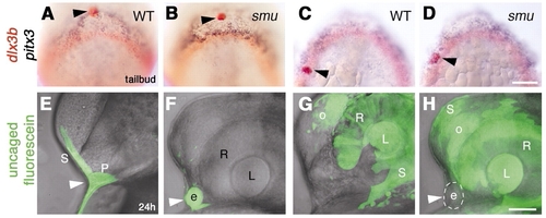
Ectopic lens formation in smoothened mutant embryos does not result from aberrant movement of lens precursors. (A-D) Bud stage embryos fixed immediately after photoactivation, then hybridized with probes for pitx3 (blue) and dlx3b (red). Photoactivated median anterior cells (A,B; n=14) and lateral posterior cells (C,D; n=11) in wild-type (A,C) and smoothened mutant (B,D) embryos labeled with anti-fluorescein (red, black arrowheads). (E-H) Embryos at prim-5 stage, after uncaging at bud stage as indicated in A-D. (E) Wild-type; skin and anterior pituitary placode are labeled (n=10; white arrowhead). (F) smoothened mutant; ectopic lens (white arrowhead) and skin (F; n=7) are labeled (photoactivation as indicated in A,B). (G,H) Wild-type (G; n=19) and smoothened mutant (H; n=7), skin, lens, retina and olfactory placode labeled (photoactivation approximately as indicated in C,D). (H) Ectopic median lens (white arrowhead, circle) is not labeled in smoothened mutant embryos (n=7). More cells were labeled in G,H than indicated in control embryos (C,D). (E-H) Superimposed bright-field images and fluorescent confocal stacks. e, ectopic lens; L, retina-associated lens; o, olfactory placode; P, pituitary; R, retina; S, skin;. (A-D) Dorsal views of prospective head region, anterior towards top; (E-H) side views, anterior towards the left, dorsal towards the top. Scale bars: 100 μm in A-D; 50 μm in E-H. |