
Fig. 1
- ID
- ZDB-FIG-050601-2
- Publication
- Oates et al., 2005 - Cooperative function of deltaC and her7 in anterior segment formation
- Other Figures
- All Figure Page
- Back to All Figure Page
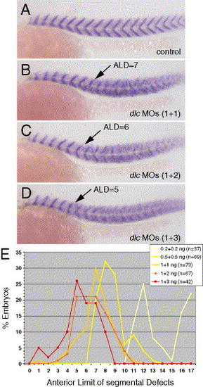
Effect of reduction of deltaC function on zebrafish segmentation. (A–D) Myotome boundaries of the trunk marked by titin expression are shown in 26 hpf embryos in lateral view with anterior to the left and dorsal upwards. Arrows in panels (B–D) indicate Anterior Limit of Defects (ALD) in each embryo. (A) Uninjected control. Representative embryos injected with 1 ng/nL (B), with 1 and 2 ng/nL (C) and with 1 and 3 ng/nL (D) of each of the morpholinos dlcMO3 and dlcMO4, respectively. (E) Data from a histogram (plotted as line graph) showing the distribution of ALD for populations of embryos injected with increasing concentrations of combined morpholinos, indicating a final ALD at segment 5. Embryos injected with doses higher than 1 + 3 ng/nL of the combined morpholinos exhibited necrosis and non-specific defects and were not included. |
| Gene: | |
|---|---|
| Fish: | |
| Knockdown Reagents: | |
| Anatomical Term: | |
| Stage: | Prim-5 |
Reprinted from Developmental Biology, 280(1), Oates, A.C., Mueller, C., and Ho, R.K., Cooperative function of deltaC and her7 in anterior segment formation, 133-149, Copyright (2005) with permission from Elsevier. Full text @ Dev. Biol.