|
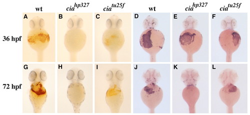
Characterization of the embryonic blood phenotype in cia. (A-L) Ventral views of the anterior region of embryos. (A-C,G-I) Whole-mount o-dianisidine staining of wild-type and cia embryos. Compared with wild type at 36 hpf (A), ciahp327 (B) (shown as representative of ciahs019 and ciaiu089 at all stages) lack hemoglobinized erythrocytes, while ciatu25f (C) manifest a moderate decrease. At 72 hpf, circulating hemoglobinized erythrocytes are still absent in ciahp327 (H) and a moderate decrease is again observed in ciatu25f (I) compared with wild type (G). (D-F,J-L) Whole-mount RNA in-situ hybridization for ße1 globin in wild-type and cia embryos. (D-F) At 36 hpf, cia embryos are indistinguishable from wild type, while the onset of anemia in cia is apparent at 72 hpf, with ciahp327 (K) possessing less than approximately 30% of cells compared with wild type (J), and ciatu25f (L) exhibiting an approximate 50% decrease in erythrocytes.
|