
Fig. 2
- ID
- ZDB-FIG-050304-1
- Publication
- Iovine et al., 2005 - Mutations in connexin43 (GJA1) perturb bone growth in zebrafish fins
- Other Figures
- All Figure Page
- Back to All Figure Page
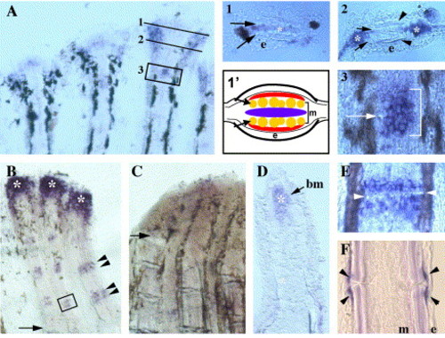
Expression of connexin 43 in fins. In situ hybridization was completed using a probe that recognizes the cx43 coding sequence. (A) Ontogenetically growing fin. Numbers indicate areas of further interest. (1) Transverse section through distal crescent. Arrows, actinotrichia; e, epidermis; *, cx43-expressing cells. (1′) Cartoon of transverse section through the distal crescent. Arrows, actinotrichia; e, epidermis; m, mesenchyme. Blue reflects undifferentiated cells medial to actinotrichia. Red represents cells which have crossed actinotrichia and become determined as osteoblasts (i.e., evx1-positive cells). (2) Transverse section through lateral cx43-stained cells. Arrows, actinotrichia; arrowheads, lepidotrichia; e, epidermis; *, cx43-expressing cells. (3) High magnification of a representative young joint, similar to boxed region in A. Arrow points to newly forming joint. Bracket identifies cx43-stained cells. (B) Wild-type regenerating fin at 5 dpa. Arrow, amputation plane; arrowheads, cx43-stained cells around mature joint; *, cx43-expressing cells in the blastema. (C) sofb123 regenerating fin at 5 dpa. Arrow, amputation plane. (D) Longitudinal cross-section through a cx43-stained regenerating fin. The basement membrane (bm) separates the epithelial and mesenchymal compartments. *, cx43-expression. (E) High magnification of a representative mature joint, similar to boxed region in B. Arrowheads point to joint. (F) Longitudinal cross-section through a mature joint. Arrowheads, cx43-stained cells; e, epidermis; m, mesenchyme. |
| Gene: | |
|---|---|
| Fish: | |
| Anatomical Term: | |
| Stage: | Days 45-89 |
Reprinted from Developmental Biology, 278(1), Iovine, M.K., Higgins, E.P., Hindes, A., Coblitz, B., and Johnson, S.L., Mutations in connexin43 (GJA1) perturb bone growth in zebrafish fins, 208-219, Copyright (2005) with permission from Elsevier. Full text @ Dev. Biol.