
Fig. 2
- ID
- ZDB-FIG-050209-12
- Publication
- Tyurina et al., 2005 - Zebrafish Gli3 functions as both an activator and a repressor in Hedgehog signaling
- Other Figures
- All Figure Page
- Back to All Figure Page
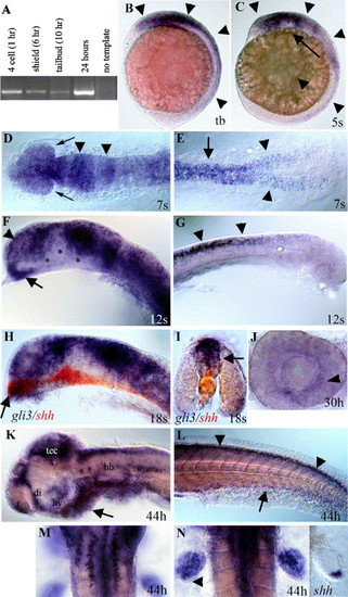
Developmental expression of zebrafish gli3. (A) RT–PCR analysis using gli3-specific primers shows that maternal gli3 mRNA is present at the four-cell stage (prior to zygotic transcription), as well as at shield, tail bud, and 24-h stages, after zygotic transcription has begun. (B) Tail bud stage (10 h). gli3 is first expressed in neural ectoderm (arrowheads) with expression most intense in the brain. (C) Five-somite stage (12 h). During early somitogenesis, gli3 is expressed throughout the brain and spinal cord (arrowheads) with expression being lost in more ventral neural tissue (arrow). (D, E) Seven-somite stage (13 h). Dorsal view. (D) gli3 is expressed throughout the embryonic brain and developing eyes (arrows) with more intense stripes of expression in the presumptive midbrain and midbrain/hindbrain border (arrowheads). (E) In the trunk, gli3 is expressed in the spinal cord (arrow) and in the lateral neural plate prior to neural tube formation (arrowheads). (F, G) Twelve-somite stage (15 h). (F) At 15 h, gli3 is expressed throughout the telencephalon (arrowhead) and anterior diencephalon (arrow) in the forebrain. (G) At 15 h, gli3 is strongly expressed in the spinal cord and is restricted to the dorsal neural tube (arrows). (H, I) Eighteen-somite stage (17 h). (H) Dorsal expression of gli3 (purple/blue) is largely complementary to the more ventral expression of shh (red) in the brain. However, in the most anterior diencephalon (arrow in H), gli3 expression overlaps with shh expression. (I) Cross-section through the spinal cord shows that gli3 (purple) is expressed in the neural tube dorsal to shh (red), which is in the floor plate (arrow) and notochord. (J) At 30 h, lateral view of dissected eye. gli3 is expressed weakly in the eye in cells surrounding the lens. (K–N) At 44 h. (K) At later stages, gli3 expression becomes restricted to the anterior telencephalon, dorsal diencephalon, hypothalamus, tectum, and dorsal hindbrain. Small clusters of cells also express gli3 in the ventral hindbrain. Mesendoderm ventral to the brain also expresses gli3 (arrow). (L) In the trunk, gli3 expression is restricted to the most dorsal regions of the spinal cord (arrowheads) and ventral endoderm (arrow). (M) Dorsal view of hindbrain showing two rows of ventral cells (see K for lateral view) on either side of the midline that express gli3. (N) gli3 is expressed throughout the fin bud with the exception of cells at the posterior margin (arrowhead). This expression overlaps with, but is much broader than, that of shh (right panel). (B, C, F–H, J–K) Side views, anterior to the left. (D, E, M, N) dorsal views. (I) Cross-section through trunk. (D–L) Yolks removed. (D, F, H) Eyes removed. Abbreviations: di; diencephalon, hb; hindbrain, hy; hypothalamus, tb; tail bud, t; telencephalon, tec; tectum. (For interpretation of the references to colour in this figure legend, the reader is referred to the Web version of this article.) |
| Genes: | |
|---|---|
| Fish: | |
| Anatomical Terms: | |
| Stage Range: | 4-cell to High-pec |
Reprinted from Developmental Biology, 277(2), Tyurina, O.V., Guner, B., Popova, E., Feng, J., Schier, A.F., Kohtz, J.D., and Karlstrom, R.O., Zebrafish Gli3 functions as both an activator and a repressor in Hedgehog signaling, 537-556, Copyright (2005) with permission from Elsevier. Full text @ Dev. Biol.