
Fig. 5
- ID
- ZDB-FIG-041208-2
- Publication
- Esni et al., 2004 - Notch inhibits Ptf1 function and acinar cell differentiation in developing mouse and zebrafish pancreas
- Other Figures
- All Figure Page
- Back to All Figure Page
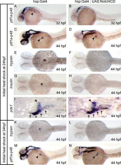
Notch pathway activation delays exocrine differentiation in developing zebrafish pancreas. Heat-shocked embryos expressing a hsp:Gal4 transgene alone or in combination with UAS:notch1aICD were assessed for initiation of ptf1a-p48 and trypsin expression, as well as expression of insulin and pdx1. Following heat shock at 24 hpf, ptf1a-p48 expression is delayed in 32 hpf hsp:Gal4;UAS:notch1aICD embryos (B) compared with hsp:Gal4 controls (A), but recovers by 34 hpf (data not shown). As assessed at 44 hpf, both trypsin (E,F) and insulin (G,H) expression are reduced in hsp:Gal4;UAS:notch1aICD embryos compared with hsp:Gal4 controls, even while ptf1a-p48 (C,D) and pdx1 (I,J) expression remain normal. Following delayed heat shock initiated at 34 hpf (after normal onset of ptf1a-p48 expression), ptf1a-p48 expression remains normal at all time points (M,N), whereas expression of trypsin is delayed (K,L). Arrowheads in A,C,D,M,N indicate endodermal domain of ptf1a-p48 expression, distinct from expression in developing hindbrain. Arrowheads in I and J indicate pdx1-positive principal islet; arrows indicate adjacent pdx1-positive intestine. |