
Anatomy of the 24, 48, 72 and 120 hours Zebrafish (Danio
rerio) Embryo
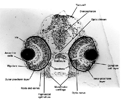
This collection of sections through zebrafish embryos at four different
stages of development is thought to provide some help to understand how
the zebrafish embryo looks inside.
Thin section in Araldite were stained with methylene blue. Images were taken
and digitized.
You will find an overview image for each stage, with links (click at numbers
on image) to images of the sections. You can also download high resolution
images (JPEG, about 1 MB), which in most cases are good enough to zoom in
down to the level of individual cell nuclei.
None of us is a classical fish biologist or did study anatomy. While we
tried hard, inevitably there will be mistakes and omissions. Please contact
Wolfgang Driever (Driever@ruf.uni-freiburg.de)
with suggestions for improvements.
Contributions:
The identification of the parts of the embryo was done by:
Salim Abdelilah, Wolfgang Driever, Alan Gorn, Jarema Malicki, Stephan Neuhauss,
Michael Pack, Zehava Rangini, Alexander Schier, Lilianna Solnica-Krezel,
Didier Stanier, Derek Stemple.
Thanks to Chuck Kimmel for providing the pictures of live embryos we use
for navigating the anatomy sections. (Kimmel, C. B., Ballard, W. W., Kimmel,
S. R., Ullmann, B. and Schilling, T. F. (1995). Stages of embryonic development
of the zebrafish. Dev. Dyn. 203, 253-310.)
Chuck Kimmel, Steve Wilson and Bill Ballard helped correct our wildest mistakes.
The sections, prints and artwork were done by:
Lisa Vogelsang, Thomas Binder, Lisa Anderson and Jane Belak.
WWW page designed by Jonathan Delgado
Sole responsibility for all the mistakes: Wolfgang Driever