
- Title
-
Rare missense variants in FNDC1 are associated with severe adolescent idiopathic scoliosis
- Authors
- Charng, W.L., Haller, G., Whittle, J., Nikolov, M., Avery, A., Morcuende, J., Giampietro, P., Raggio, C., Miller, N., Justice, A.E., Strande, N.T., Seeley, M., Bodian, D.L., Wise, C.A., Sepich, D.S., Dobbs, M.B., Gurnett, C.A.
- Source
- Full text @ J. Med. Genet.
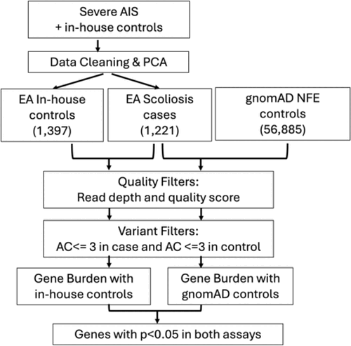
Flow chart of gene burden analysis. Exomes of severe adolescent idiopathic scoliosis (AIS) cases and in-house controls went through data cleaning and anchoring to European population. Rare gene burden analyses were performed between 1221 unrelated severe AIS probands with two independent controls (1397 in-house controls and 56885 gnomAD non-Finnish European (NFE) controls) in the autosomal dominant mode using the Testing Rare vAriants using Public Data (TRAPD) method. PCA, principal component analysis. AC, allele count. EA, European American. |

Localisation and segregation of FNDC1 variants. Rare FNDC1 missense variants are enriched in AIS cases and segregate with incomplete penetrance in AIS families. (A) Segregation of rare FNDC1 missense variants in severe AIS families from the gene burden analysis. (B) Segregation of FNDC1 variants in additional AIS families not included in the original gene burden analysis. (C) FNDC1 variants tested in segregation analyses in this study. (D) Rare FNDC1 missense variants observed in severe AIS cases and in-house controls. Blue bars represent fibronectin type III domains. Variants enriched in severe AIS cases are marked in red and variants enriched in controls are marked in blue. Variants with equal allele count in cases and controls are marked in grey. AIS, adolescent idiopathic scoliosis; FNDC1, fibronectin type III domain containing 1. |

The fndc1 nonsense allele (fndc1 sa23734) is associated with increased bone mineral density and total vertebral cross-sectional area in zebrafish. (A) MicroCT scan images demonstrating 3D projections of the vertebra of an individual fish. Vertebra in a lateral plane (top left) and axial plane (bottom left). Total vertebral cross-sectional area is quantified by determining the area of each axial section (right). MicroCT scans of zebrafish (n=5 for each genotype) revealed that fndc1 mutants have (B) increased vertebral bone mineral density (fndc1 +/+ vs fndc1 +/sa23734: p=0.034; fndc1 +/sa23734 vs fndc1 sa23734/sa23734: p=0.00019; fndc1 +/+ vs fndc1 sa23734/sa23734: p=6.08E-06) and (C) increased vertebral cross-sectional area compared with wild-type siblings (fndc1 +/+ vs fndc1 +/sa23734: p=0.0011; fndc1 +/sa23734 vs fndc1 sa23734/sa23734: p=0.94; fndc1 +/+ vs fndc1 sa23734/sa23734: p=1.37E-05). P values were calculated using the Wilcoxon rank-sum test, two sided. * indicates p< 0.05, ** for p < 0.005, *** for p<0.0005, and **** for p < 0.00005. PHENOTYPE:
|