FIGURE SUMMARY
Title

Understanding the development of tuberculous granulomas: insights into host protection and pathogenesis, a review in humans and animals

Authors
Lyu, J., Narum, D.E., Baldwin, S.L., Larsen, S.E., Bai, X., Griffith, D.E., Dartois, V., Naidoo, T., Steyn, A.J.C., Coler, R.N., Chan, E.D.
Source
Full text @ Front Immunol

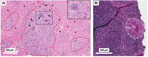
Human TB granulomas may be non-necrotic or necrotic. Several different fates of granulomas occur and such heterogeneity may present in the same individual. (A) A non-necrotic TB granuloma is characterized by the presence of epithelioid macrophages and activated macrophages as well as fewer foamy macrophages, necrotic macrophages, neutrophils, and more or less a rim of host-protective T cells and B cells along with relatively few bacilli. iBALT may be considered an “appendage” of granulomas typically comprised of dendritic cells, B cells, and T cells and may serve as a ready supply of immune cells for the neighboring granuloma. This granuloma is protective as shown by the relatively few Mtb (red-colored rod-shaped structures. (B) A granuloma with central necrosis characterized by fewer epithelioid and activated macrophages and more foamy macrophages, necrotic macrophages, and neutrophils along with an area of central necrosis. There are also fewer host-protective T cells and B cells. Due to the fewer number of host-protective innate and adaptive immune cells, there is overall a greater Mtb burden. However, in the central necrotic area of this human granuloma, there is active killing of Mtb resulting in fewer viable Mtb in this region. Higher levels of MmpL7 and IDO-1 inhibit iBALT formation. IDO-1, indoleamine 2,3-dioxygenase; iBALT, inducible bronchus-associated lymphoid tissue; MmpL7, Mycobacterial membrane protein Large 7; Mtb, Mycobacterium tuberculosis; TB, tuberculosis.

Peritoneal TB with non-necrotizing granulomas. A 51-year-old man from the Philippines presented with a two-year history of early satiety, intractable abdominal pain, and weight loss exceeding 60 pounds. Abdominal CT showed evidence of “peritoneal carcinomatosis.” (A) Peritoneal biopsy showed no evidence of malignancy. Instead, there were multiple non-necrotizing granulomas (demarcated by red dashed lines) with prominent Langhans-type multinucleated giant cells (arrows) and surrounding fibrosis (asterisks) (H&E). The boxed inset shows a magnified view of the smaller box demonstrating the multinucleated giant cells. (B) A peritoneal lymph node biopsy showed similar non-necrotizing granulomatous inflammation (white dashed line) with a conspicuous multinucleated giant cells (white arrow) (H&E). The acid-fast stain in both the peritoneal and lymph node biopsies were negative. With standard treatment for drug-susceptible TB, his symptoms abated and he regained his lost body weight. TB, tuberculosis.

Formation of foamy macrophages and their role in TB pathogenesis. Foamy macrophages play a key role in the formation of caseous necrosis. They are formed when macrophages acquire cholesterol-containing LDL particles into lipid bodies. See text for discussion. ESAT-6, 6 kDa early secretory antigenic target; GPR109A, a specific G-protein coupled receptor; LB, lipid bodies; LDL, low density lipoprotein; MA, mycolic acids; PPARγ, peroxisome proliferator-gamma; TR4, testicular receiver 4.

Mechanisms by which deficiency or excess CD4+IFNγ+ T cells predispose to TB. (A) Deficiency of TH1 (CD4+IFNγ+) T cells – as seen in advanced AIDS, genetic defects in the IFNγ-IL-12 axis, and in animal models – has been shown to increase the risk of TB and other mycobacterial infections. This IFNγ deficiency leads to an increase in IL-17 and neutrophilic influx. This combined with a lack of macrophage activation (due to insufficient number of CD4+IFNγ+ T cells) leads to an overwhelming Mtb infection of phagocytes, resulting in an unprotective, necrotic granuloma. (B) Excess TH1 cell activation and numbers lead to a secondary increase in TNF, which can then induce RIP1 and RIP3 activation, excessive ROS formation, and macrophage necroptosis. The sequence of these events leads to over-inflamed, necrotic, and unprotective granulomas. (C) There is ample evidence in both humans and experimental animal models that optimal quantity and temporal influx of Mtb-specific CD4+IFNγ+ T cells are necessary for control ± eradication of Mtb infection, in part through activation of macrophages. IFNγ also inhibits IL-17 production from TH17 cells which can induce chemokines for CD4+IFNγ+ T cells. In non-hematopoietic cells, IFNγ also induces IDO-1 production that inhibit excessive IL-17 production (via the catabolized products of tryptophan by the actions of IDO). Hence, IL-17 production is kept in check, limiting the amount of potentially harmful neutrophilic influx. IDO-1, indoleamine 2,3-dioxygenase; IFNγ, interferon-gamma; IL-12, interleukin-12; IL-17, interleukin-17; Mtb, Mycobacterium tuberculosis; RIP, receptor-interacting serine-threonine kinases; ROS, reactive oxygen species; TB, tuberculosis; TNF, tumor necrosis factor.

Differential expression of hypoxia, HIF-1α, and downstream effects in the central and peripheral regions of the necrotic granuloma. The central region of a necrotic granuloma is more hypoxic. As a result, there is greater HIF-1α expression with a panoply of downstream effects due to HIF-1 activity. One immune mechanism induced by HIF-1 is aerobic glycolysis, which in turn induces autophagy through the byproduct lactate. Hence, the central necrotic area of an intact human TB granuloma is usually sterile or with low bacterial burden. In contrast, the peripheral region of the granuloma less hypoxic with less HIF-1α induction. In the presence of macrophages that are less effective in killing Mtb (as shown by the foamy macrophages), there is less ability to kill Mtb, as shown by the greater bacilli number. However, in the presence of activated macrophages and T cells, the peripheral granuloma may also be efficient in either killing or controlling the Mtb infection. HIF-1α, hypoxia-inducible factor 1-alpha; IFNγ, interferon-gamma; Mtb, Mycobacterium tuberculosis; TB=tuberculosis;.

Either excessive or deficiency of molecular or cellular immune elements may predispose to unprotective granuloma or defense against Mtb. The diagram illustrates that either deficiency or excessive of specific immune components (x-axis) may result in increased susceptibility and severity of TB (y-axis). Using the immune checkpoint inhibitors as an example, we posit that in conditions where the host is either severely immunocompromised, immune checkpoint inhibition would be beneficial against mycobacterial infection, as exemplified by: (i) advanced AIDS patient with severe mycobacterial infection that improved with an anti-PD-1 antibody and (ii) TIM3 antagonism or TIM3 gene knockout mice are protected against mycobacteria in the less immunocompetent BALB/c mice. In contrast, in hosts with better preserved immune function, immune checkpoint inhibition results in a hyperinflammatory state, worsening control of mycobacterial infections, as exemplified by the mice with genetic disruption for PD-1 or PD-L1 gene. Perhaps the severity of TB is not as severe in the PD-L1 knockout mice than the PD-1 knockouts because in the PD-L1 knockout mice, PD-L2 is still intact and thus the enhanced immune activation with the genetic disruption of PD-L1 is not as severe as of PD-1. We have also shown other molecular and cellular components in which deficiency or excess predispose to TB. See also text and Table 2 for further discussion. AIDS, acquired immunodeficiency syndrome; HIF-1α, hypoxia-inducible factor 1-alpha; HO-1, heme oxygenase-1; ICI, immune checkpoint inhibitor; IDO-1, indoleamine 2,3-dioxygenase; IFNγ, interferon-gamma; IL-12, interleukin-12; IL-17, interleukin-17; LTA4H, leukotriene A4 hydrolase; MAC, Mycobacterium avium complex; MP, macrophages; MMP, matrix Mtb, Mycobacterium tuberculosis; NFκB, nuclear factor-kappa B; PD-1, programmed cell death protein-1; PD-L1, programmed death-ligand-1; TB, tuberculosis; TGFβ, transforming growth factor-beta; TNF, tumor necrosis factor.

Relative IDO-1 activity helps dictate the level of protection by TB granulomas. IDO-1 activity can also be an indicator of the protectiveness of a granuloma. When there is an optimal level of IDO-1 inhibition, the remodeling of the granulomas allows access of CD4+ T cells to the central core of the granuloma, resulting in Mtb death. However, excessive or insufficient IDO-1 activity leads to an unprotective granuloma. An excess of IDO activity results in an unprotective, hypoinflammatory granuloma, characterized by deficient proliferation of CD4+ and CD8+ T cells and poor access of CD4+ T cells to the granuloma core, allowing Mtb to grow within the center. On the other hand, an insufficient level of IDO-1 activity leads to an unprotective, hyperinflammatory granuloma, characterized by excess IL-17 production and excess neutrophil influx and granuloma necrosis. IDO-1 is also responsible for the catabolization of tryptophan (a molecule necessary for Mtb growth) into kynurenine. A deficiency of IDO-1 would in turn increase levels of tryptophan and allow for more Mtb growth. IDO-1, indoleamine 2,3-dioxygenase; IL-17, interleukin-17; Mtb, Mycobacterium tuberculosis;.

Origin of Mtb and early events in the development of post-primary TB. (A) An often-cited description for the evolution of post-primary (reactivation) TB is that immune dysfunction results in progressive necrosis and liquefaction of the granuloma, exuberant multiplication of dormant Mtb, disruption of the integrity of the granuloma wall, and spillage Mtb-filled necrotic granulomatous content into the airways. Pneumonia forms due to endobronchial spread of granuloma content. (B) An alternative description of post-primary TB is based on the paradigm chronic stable granulomas are mostly sterile and that the dormant Mtb that “reawaken” originated from lymph nodes. fat cells, and airway epithelial cells causing a bronchogenic lipoid pneumonia. Granulomas may form around pockets of necrotizing pneumonia. As discussed in the text, it is plausible that both reactivation models occur. Mtb, Mycobacterium tuberculosis; TB, tuberculosis.

Distribution of lesions in primary and post primary TB. The localization of lesions of (A) primary and (B) post-primary TB of several people were plotted on an X-ray. Each dot marks the location of an individual’s lesion. The distribution of primary TB is consistent with chance distribution of air-borne infection while most post-primary lesions were in the upper lobes, especially the apical segments. Reprinted with permission of the American Thoracic Society. Copyright © 2024 American Thoracic Society. All rights reserved. Medlar E.M. The pathogenesis of minimal pulmonary tuberculosis: A study of 1,225 necropsies in cases of sudden and unexpected death. Am Rev Tuberc 1948; 58: 583-611. The American Journal of Respiratory and Critical Care Medicine is an official journal of the American Thoracic Society. TB, tuberculosis.

Anatomical-physiological-chemical mechanisms by which post-primary TB is more common in the upper lobes of the lungs. Based on differences in the physiological-chemical properties between the upper lobes and lower lobes, primary Mtb lesions located in the upper lobes, while fewer than that in the lower lobes, are more likely to reactivate. (A) Diagram showing that primary TB is located mostly in the lower lung zone (green circular structure that represents a calcified granuloma – Ghon complex). This distribution of primary TB is due to greater (3.4X) ventilation in the lower lobes than the upper lobes of an upright human. (B) In the upright human, there is decreased lung perfusion pressure due to lower pulmonary artery pressure in the upper lobes. This decreased perfusion will result in decreased influx of immune cells and soluble mediators to the upper lobes in the upright position. (C) Lymph formation and flow are also significantly decreased in the upper lobes, resulting in increased accumulation of mycobacteria and antigens in the upper lobes and contributing to increased hypersensitivity reactions and lung damage. pH in the upper lobes is more alkalotic which induces glycolysis and, in turn, increases autophagic clearance of Mtb by the macrophages. (D) The PAO2 is also significantly greater in the upper lobes than the lower lobes, which favors Mtb growth. The mechanical stress and alveolar size of the upper lobes are also greater (by 40X and 4X, respectively) than the lower lobes, predisposing this area to the development of cavities, especially with large inspiratory efforts. Mtb, Mycobacterium tuberculosis; PAO2, partial pressure of alveolar oxygen; TB, tuberculosis.

Different types of TB lung lesions in the C3HeB/FeJ mice. Three types of histopathologic TB lesions are seen in the C3HeB/FeJ mice. (A) Type 1 lesion is mainly comprised of foamy macrophages, few epithelioid macrophages and lymphocytes, and a caseous center surrounded by neutrophils. This lesion most resembles to that seen in human TB. (B) Type 2 lesion is the most severe form and is characterized by a neutrophilic, necrotizing alveolitis with occlusion of the terminal bronchioles and caseous necrosis. (C) Type 3 lesion is comprised mainly of infiltration of epithelioid macrophages and lymphocytes, smaller numbers of foamy and activated macrophages and neutrophils around the airway and blood vessel pair. This is the typical lesion seen in most other mouse strains. TB, tuberculosis.

Acknowledgments
This image is the copyrighted work of the attributed author or publisher, and ZFIN has permission only to display this image to its users. Additional permissions should be obtained from the applicable author or publisher of the image. Full text @ Front Immunol