FIGURE SUMMARY
Title

RPTOR blockade suppresses brain metastases of NSCLC by interfering the ceramide metabolism via hijacking YY1 binding

Authors
Lin, Y., Wu, Y., Zhang, Q., Tu, X., Chen, S., Pan, J., Xu, N., Lin, M., She, P., Niu, G., Chen, Y., Li, H.
Source
Full text @ J. Exp. Clin. Cancer Res.

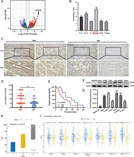
Association of high RPTOR expression or the ceramide pathway with NSCLC-BM. A, B RNA sequencing showed that RPTOR was more highly expressed in BM than other genes like PLP1, GFAP, TF, PMP2 and TUBB4A, which was validated by qRT-PCR. C, D IHC analysis showed that the BM + group reported a higher RPTOR expression and score than the BM- group (100x, 200 × magnification, scale bar = 100 μm). E Kaplan–Meier analysis showed that the median survival was significantly lower in the high RPTOR (18 samples with IHC scores ≥ 6) than in the low RPTOR subgroup (15 samples with IHCs ≤ 3) in the BM + patients. F, G H1299 and PC9 cell lines were chosen to be our experiment cell lines due to their significantly higher RPTOR expression than those in other LUAD cell lines. H, I The whole-exome sequencing (WES) was used to screen different genes from the BM + and BM- groups. A DAGM model was constructed to assess the role of somatic variants in the signaling pathways. The ceramide signaling pathway was found to be involved in the BM progress

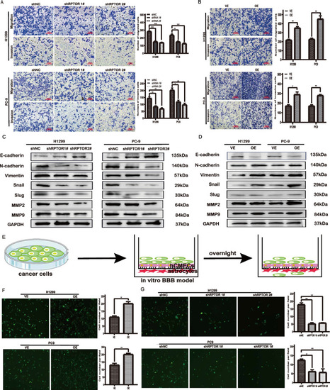
The RPTOR-promoted traverse of lung cancer cells to the BBB. A, B Transwell assays showed that RPTOR promoted the migration and invasion of lung cancer cell lines. C, D Western blotting reported that RPTOR increased EMT-related proteins and extracellular matrix. E, F, G Fluorescence microscope transwell assays showed that RPTOR promoted the traverse of lung cancer cells to the BBB

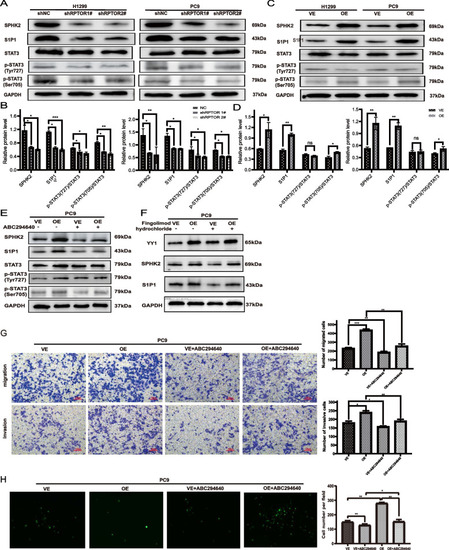
The RPTOR-promoted BM of NSCLC through the SPHK2/S1P/STAT3 signaling pathway. A-D Western blotting showed that the exogenous RRTOR boosted the expression of proteins of the ceramide metabolism pathway, including SPHK2, S1PR1 and p-STAT3 (ser705). EH The SPHK2 inhibitor, ABC294640, reversed the activation of the SPHK2/S1P/STAT3 signaling pathway and the RPTOR overexpression-increased cell invasiveness and permeability of BBB, while the S1P inhibitor, fingolimod hydrochloride, reduced the expression of S1P1 protein, without affecting the expression of YY1 and SPHK2

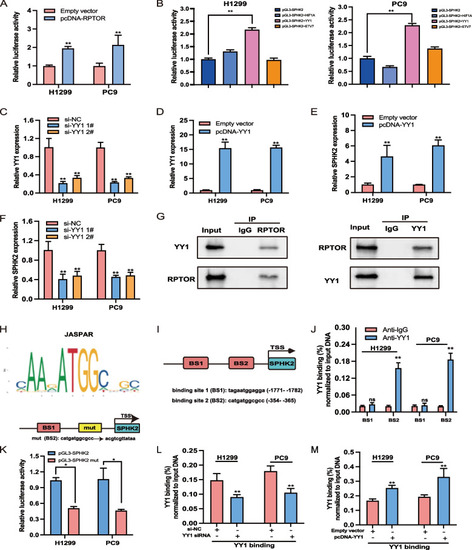
The RPTOR-regulated SPHK2/S1P pathway via binding to transcription factor YY1. A Dual-luciferase reporter assays indicated that RPTOR enhanced SPHK2 expression in H1299 and PC9 cell lines. B Transcription factor YY1 was screened to significantly enhance the transcriptional activity of SPHK2 when compared with other candidate transcription factors like HIF1A and ETV7, when co-transfected with pGL3-SPHK2. C-F qRT-PCR assays revealed a positive correlation between SPHK2 and YY1 expression, with both YY1 and SPHK2 expression down-regulated after treatmenting PC9 and H1299 cells with pcDNA-YY1 or YY1 siRNA. G Co-IP assays showed mutual interactions between RPTOR and YY1 in lung cancer cell lines. HK The JASPAR database was used to predict YY1-binding sites in the SPHK2 promoter sequences. BS1 and BS2 were predicted to be YY1-binding sites, with BS2 at -353 ~ -365 nt of SPHK2 reporting a greater DNA enrichment by ChIP assays. The enrichment of SPHK2 DNA was reduced by mutation of BS2, confirming that BS2 is the site in the SPHK2 promoter bound by YY1. L-M Effect of YY1 expression on SPHK2 promoter sequences. YY1 enriched fewer SPHK2 promoter sequences after si-YY1 1# transfection but more sequences after pcDNA-YY1 transfection

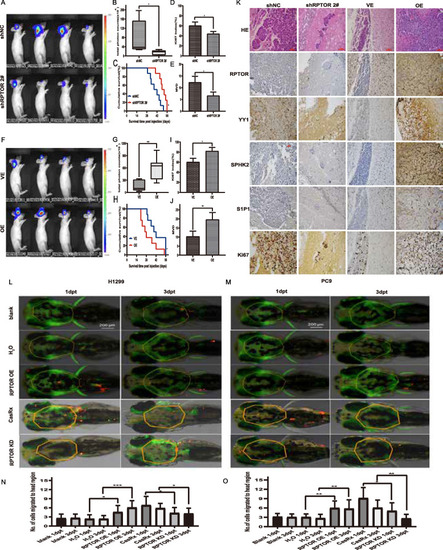
The suppressed BM of NSCLC and attenuated SPHK2/S1P/STAT3 pathway in xenograft and zebrafish model by RPTOR blockade. A-J PC-9 cells were injected intravenously to construct the BM mouse model. Fluorescence microscopy showed that RPTOR blockade significantly suppressed the BM formation, the fluorescence of tumor cells in the ROI of BM, the expression of the proliferation marker Ki67 and MVD in the peritumoral area, prolonging overall survival (200 × magnification, scale bar = 50 μm). RPTOR overexpression produced the opposite effects. K Effects of RPTOR expression on the expression of proteins in BM tissue samples in nude mice. RPTOR knockdown reduced the expressions of RPTOR, YY1, SPHK2, and S1P1, with RPTOR overexpression yielding the opposite effects (200 × magnification, scale bar = 50 μm). L-O H1299 and PC9 cells bearing vectors for RPTOR knockdown or overexpression were injected into the perivitelline space of zebrafish embryos. The number of cancer cells at days 1 and 3 after the injection was significantly higher in the RPTOR-overexpressed group than in their respective control groups, whereas the number of cancer cells at days 1 and 3 after injection was significantly lower in RPTOR-knockdown cells than in control cells

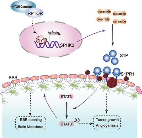
The mechanism underlying the RPTOR-promoted NSCLC-BM via the SPHK2/S1P/STAT3 signaling pathway. RPTOR regulated the transcriptional activation of the ceramidase SPHK2 by binding to the transcription factor YY1. SPHK2 catalyzed the degradation of ceramide to S1P. The latter, in turn, activated the STAT3 signaling pathway by binding to S1PR1 (solid lines), altering the microenvironment and enhancing the permeability of the BBB (dotted lines), resulting in the BM of NSCLC

Acknowledgments
This image is the copyrighted work of the attributed author or publisher, and ZFIN has permission only to display this image to its users. Additional permissions should be obtained from the applicable author or publisher of the image. Full text @ J. Exp. Clin. Cancer Res.