
- Title
-
Modelling human lower urinary tract malformations in zebrafish
- Authors
- Kolvenbach, C.M., Dworschak, G.C., Rieke, J.M., Woolf, A.S., Reutter, H., Odermatt, B., Hilger, A.C.
- Source
- Full text @ Mol Cell Pediatr
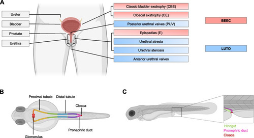
Schematic overview of human and zebrafish urinary tract. A Schematics of human urinary system. Topological subdivisions of the phenotypic complexity of BEEC (red) and LUTO (blue) are depicted. B Overview of zebrafish urinary system. It is composed of 2 nephrons with a pair of glomeruli (bright red) and tubules that can be divided in a proximal and distal segment. The proximal part is subdivided into neck (orange), proximal convoluted (yellow) and proximal straight tubule (green). The distal portion is divided in distal early (blue), corpuscle of Stannius (brown) and distal late (purple). The last segment depicts the pronephric duct (pink) that distally fuses with the cloaca (dark red). C Schematic depiction of cloacal region at 120 hpf. The hindgut (green) opens to the exterior, adjacent to the pronephric duct (pink), both fusing with the cloaca (dark red). Created with BioRender.com |
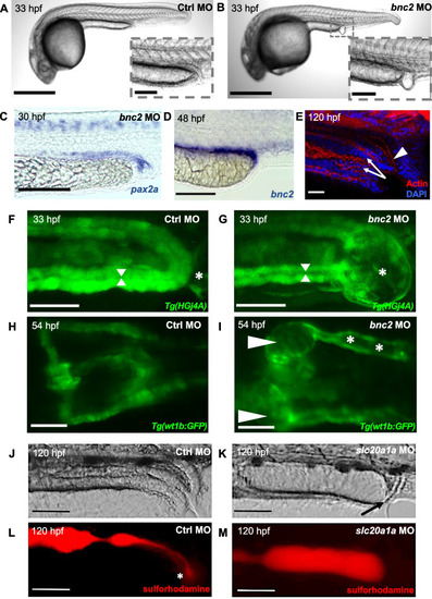
Zebrafish as a model for developmental lower urinary tract defects. A, B Zebrafish injected with bnc2 Morpholino (MO) frequently develop a pronephric distal outlet obstruction (‘vesicle’; enlargement in B) at 33 hpf compared with controls. C WISH with a pax2a probe relates the bnc2 MO induced pronephric outlet obstruction to distal parts of the pronephric ducts and the cloaca. D At 48 hpf bnc2 was expressed in the terminal section of the pronephric ducts. E Staining for actin (red) and DAPI (blue) allows the visualization of the hindgut (white arrow) and distal pronephric duct (white arrow head) in wild-type zebrafish larvae. F, G Tg(HGj4A) zfl in dorsal view at 33 hpf showing distal pronephric outlet obstruction (asterix) with resulting dilatation of pronephric ducts (indicated by arrow heads) in bnc2 MO zfl (G) compared to zfl injected with control (Ctrl) MO. H, I Proximal pronephric region of Tg(wt1b:GFP) in dorsal view with cystic dilation of glomeruli (arrow heads) and pronephric ducts (asterisks) in bnc2 MO zfl, which recapitulate human hydronephrosis (I). J–M Zfl are depicted after the ingestion of sulforhodamine. Pictures are shown in brightfield (J, K) and fluorescent red channel (L, M). The intestine and cloaca appear regular and ensure excretion of the red dye (asterisk in L) in controls. slc20a1a knockdown impairs excretory function of the cloaca (black arrow in K) and leads to distension of the hindgut. Scale bars: A, B 500 μm (100 μm magnification in B), C, D; F–I 100 μm, E 30 μm, J–M: 50 μm. A–C; F–I Modified from Kolvenbach et al. [15]. J–M Modified from Rieke et al. [17] |