FIGURE SUMMARY
Title

Zebrafish patient-derived xenograft models predict lymph node involvement and treatment outcome in non-small cell lung cancer

Authors
Ali, Z., Vildevall, M., Rodriguez, G.V., Tandiono, D., Vamvakaris, I., Evangelou, G., Lolas, G., Syrigos, K.N., Villanueva, A., Wick, M., Omar, S., Erkstam, A., Schueler, J., Fahlgren, A., Jensen, L.D.
Source
Full text @ J. Exp. Clin. Cancer Res.

ZTX models can be established directly from patient samples and accurately predict patient treatment outcomes. A,B Quantification of the relative treatment-induced tumor regression of tumors established from 9 PDX models calculated as the change in tumor size between day 3 and 0 after implantation, in groups treated with Erlotinib 10 mg/L (A) or Paclitaxel 20 mg/L (B), divided by the change in tumor size in the control group. n = 13–19. Gray bars indicate models from patients that were not treated with the indicated drug and red bars indicate models from patients that had progressed following treatment with the indicated drug. *: p < 0.05. C Quantification of the average number of metastasized cells at three days after implantation for each of the 9 validation models. n = 13–19. Green bar indicates a model from a patient with a localized cancer, red bars indicate models from patients with disseminated cancer and grey bars indicate models from patients with unknown lymph node or metastatic status. Black dashed line indicates the cut-off level for prediction of disseminated disease by the ZTX model. D H&E micrographs of tumor sections from the four patients included in this study. E-H Quantification of the relative growth (E), average number of metastasized cells (F), and relative Erlotinib- or Paclitaxel-induced tumor regression (G and H respectively), calculated as in A/B, for the four patients included in this study. Dashed line indicates the cut-off level for predicting disseminated disease. n = 14–20

Robust implantation and growth of NSCLC PDX models in zebrafish larvae. A Cartoon illustrating the tissue handling, implantation and visualization of tissue fragments or cell suspensions from mouse PDX material into zebrafish larvae. B Representative fluorescent micrographs of DiI-labeled xenografts (shown in red) generated from cell suspensions (top row) or tissue fragments (lower row), imaged immediately and three days after implantation (left and right columns respectively). C Quantifications of changes in tumor size (rel. size) between day three and zero for three separate models (M16, M5 and M3) that were implanted either as fragments or as cell suspensions in separate cohorts of zebrafish larvae. n = 18, 15, 8 larvae in the cell suspension groups and 17, 18, 14 larvae in the fragments groups respectively. *** = p < 0.001. D Quantifications of changes in tumor size (rel. size) between day three and zero for the 25 models that was implantable in the zebrafish larvae. Average size-change was 64% for all models combined. n = 12–33. E,F Quantification of changes in tumor size (rel. size) between day three and zero plotted against the viability (E) or number of cells extracted from the fragments (F). P > 0.05. G Quantification of changes in tumor size (rel. size) between day three and zero for 11 models run at two different times. Red line indicates the average of the 11 models. NS: non-significant. H-J: Quantifications of changes in tumor size (rel. size) in zebrafish larvae between day three and zero plotted against the tumor doubling time (H), take rate (I) or stromal content (J) when growing subcutaneously in mice. P > 0.05

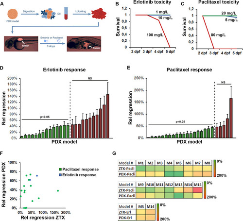
Similar response to erlotinib and paclitaxel are seen in ZTX and mouse PDX models. A Cartoon illustrating the tissue handling, implantation, and visualization of tissue fragments from mouse PDX material into zebrafish larvae followed by 3-days treatment with Erlotinib or Paclitaxel added to the water. B-C Quantification of the proportion of surviving embryos following treatment starting at 2-days post fertilization with the indicated concentrations of Erlotinib (B) or Paclitaxel (C) for one, two or three days. n = 20 (B) and n = 18 (C) per group. Blue lines indicate 1 and 5 mg/L, green lines indicate 10 and 20 mg/L and red lines indicate 100 and 80 mg/L in B and C respectively. D-E Quantification of the changes in tumor size between day three and zero for larvae treated with either Erlotinib, 10 mg/L (D) or Paclitaxel, 20 mg/L (E), divided by the changes in tumor size between day three and zero for larvae in the corresponding control groups (Rel regression) for each of the 25 models tested. Models where treatment led to significant tumor regression are shown in green whereas models where the drug did not significantly induced regression is shown in red. n = 7–22 per group, NS: non-significant. F Quantification and correlation of the treatment outcome for Erlotinib (blue dots) or Paclitaxel (green dots) plotted as relative treatment-induced regression in mouse-PDX models against that of the relative treatment-induced regression in the corresponding ZTX models. G Heat map of the degree of relative regression as quantified in D and E and shown in F for zebrafish tumor xenograft (ZTX) models and in a similar manner when PDX models grew in mice PDX, for each of the 15 models in which drug efficacy was tested in both zebrafish and mice. Erl: Erlotinib, Pacli: Paclitaxel

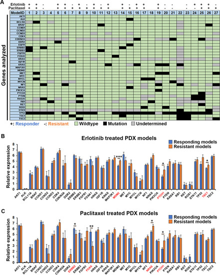
Genetics of tumors and correlation with treatment outcome. A Scheme showing the genotype of the 37 genes included in this study against the 27 PDX models analyzed. All types of mutations were combined and indicated as black squares, whereas the absence of a mutation is indicated as wildtype in green. Where low coverage did not allow an accurate assessment, this is indicated in grey. Response or lack of response to Erlotinib or Paclitaxel in the ZTX models is indicated with + or – respectively above the scheme. B Average relative expression of the 36 genes included in this study (EGFR is shown in Supplemental Fig. S2) as evaluated by RNA sequencing, for models responding (n = 14 models, blue bars) or resistant (n = 9 models, orange bars) to Erlotinib. *:p < 0.05, ***:p < 0.001. C Average relative expression of the 36 genes included in this study (EGFR is shown in Supplemental Fig. S2) as evaluated by RNA sequencing, for models responding (n = 18 models, blue bars) or resistant (n = 5 models, orange bars) to Paclitaxel. *:p < 0.05, **:p < 0.01

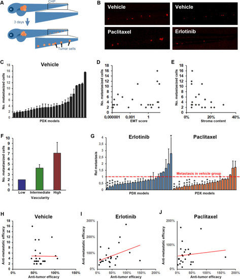
Tumor dissemination correlates with vascularity of the tumor but not tumor growth/regression rate. A Cartoon illustrating dissemination of tumor cells (shown in orange) to the caudal hematopoietic plexus (marked by the black squares) three days following tumor implantation in the perivitelline space. B Representative images of tumor cells (shown in red) in the caudal hematopoietic plexus three days after tumor implantation for a representative model in which dissemination was moderately but significantly inhibited by treatment with Erlotinib (10 mg/L, right panels) but not by treatment with Paclitaxel (20 mg/L, left panels). C Quantification of the average number of cells that at three days post implantation have disseminated to the caudal hematopoietic plexus (metastasized cells) for each of the implantable models. D-F Quantification of the EMT-score (D), stromal content (E) and vascularity (F) of the PDX models when grown in mice plotted against the average number of metastasized cells, as quantified in C, three days after implantation of the models in zebrafish larvae. Positive correlation was significant for metastasis and vascularity but not for metastasis and EMT-score or stromal content. n = 12–33 per group. G Quantification of the relative change in metastasis to the caudal hematopoietic plexus in treatment compared to vehicle groups after treatment with either Erlotinib (10 mg/L, shown in light blue), or Paclitaxel (20 mg/L, shown in orange). Red dashed line indicates the control group for each model. n = 7–22 per group. H-J Quantification of the changes in tumor size between day three and zero for larvae in the control group (H) or after treatment with Erlotinib, 10 mg/L (I), or Paclitaxel, 20 mg/L (J), divided by the changes in tumor size between day three and zero for larvae in the corresponding control groups (anti-tumor efficacy) and plotted against the relative change in metastasis as quantified in G, for each of the 25 implantable models. A non-significant trend towards a positive correlation was observed in the Erlotinib treated group but not in the Paclitaxel treated groups. n = 7–22 per group

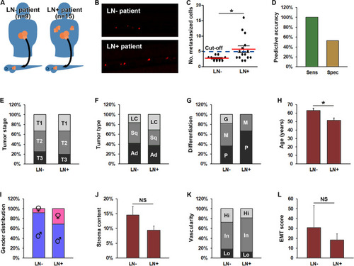
Increased metastasis in ZTX models accurately predicts lymph node involvement in the patients. A Cartoon illustrating implantation of PDX models generated from non-small cell lung cancer patients with or without lymph node involvement (LN+, right part of the figure and LN-, left part of the figure respectively) in zebrafish larvae. B Representative fluorescent micrographs of tumor cells (shown in red) disseminated to the caudal hematopoietic plexus three days after implantation from patients having lymph node negative (LN-) or positive (LN+) disease upon diagnosis. C Quantification of the average number of tumor cells disseminated to the caudal hematopoietic plexus (metastasized cells) three days following implantation of PDX models generated from patients with lymph node negative (LN-) or positive (LN+) disease. Dashed line indicates the establish diagnostic cut-off of 5 disseminated cells. *: p < 0.05. n = 12–33 larvae per model. D Quantification of the diagnostic sensitivity (accuracy of predicting patients having lymph node positive disease) and selectivity (accuracy of predicting patients not having lymph node positive disease) using the cut-off value shown in C. n = 7 and 17 for sensitivity and specificity respectively. E Distribution of tumor stages associated with lymph node negative (LN-) or positive (LN+) disease. No stage was significantly associated with lymph node involvement. The number of patients associated with each group is given in Supplemental Table 1. F-I distribution of histological tumor sub-types (F), tumor differentiation stages (G), average age (H), and gender (I), among patients with lymph node negative (LN-) or positive (LN+) disease upon diagnosis. Female gender, younger age, large cell carcinoma and poorly differentiated cancer were significantly associated with higher risk of lymph node positive disease (p < 0.05). The number of patients in each group is given in Supplemental Table 1. LC: Large cell carcinoma. Sq: Squamous cell carcinoma. Ad: Adenocarcinoma. J-L Quantification of the average stromal content (J), vascularity (K) and EMT-score (L) of PDX models grown in mice derived from patients with either lymph node negative (LN-) or positive (LN+) disease upon diagnosis. n-values for each group are given in A. NS: non-significant. G: Good. M: Moderate. P: Poor. Hi: High. In: Intermediate. Lo: Low

Acknowledgments
This image is the copyrighted work of the attributed author or publisher, and ZFIN has permission only to display this image to its users. Additional permissions should be obtained from the applicable author or publisher of the image. Full text @ J. Exp. Clin. Cancer Res.