
- Title
-
Evidence of regional specializations in regenerated zebrafish retina
- Authors
- Stenkamp, D.L., Viall, D.D., Mitchell, D.M.
- Source
- Full text @ Exp. Eye. Res.
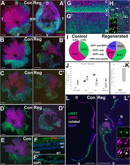
Fig. 1. Regionalized patterning of LWS1 and LWS2 cones in control and regenerated retinas of adult zebrafish at 21 days following ouabain lesion. A-D. Left whole retinas (A,B,C,D; undamaged controls; Con) and contralateral right whole retinas (A′,B′,C′,D’; regenerated; Reg) of lws:PAC(H) zebrafish, in which lws1 is reported by GFP (LWS1 cones; green) and lws2 is reported by RFP (LWS2 cones; magenta), counterstained with DAPI (blue). D, dorsal; N, nasal; V, ventral; T, temporal. In each preparation, LWS1 cones populate ventral retina and portions of peripheral retina, generally nasal, while LWS2 cones populate central and dorsal retina. Horizontal rectangle in panel B is region enlarged in panel G; vertical rectangle in panel B is region rotated and enlarged in panel G’; vertical rectangle in panel D′ is region enlarged in panel H. E. Additional undamaged control retina displaying characteristic patterns of LWS1 and LWS2 cones. F. Cryosections of undamaged control retina (F) and contralateral damaged retina (F′), obtained at four days post-injury (4DPI), stained with ZPR1 antibody (green fluorescence), which labels all LWS and RH2 cones, transgenic for sws2:mCherry, which reveals SWS2 cones (red fluorescence), counterstained with DAPI (blue); onl, outer nuclear layer; inl, inner nuclear layer; gcl, ganglion cell layer. The 4DPI damaged retina in F′ shows that the retinal layers are not intact, but retina contains DAPI + nuclei and debris of photoreceptors. Photoreceptor morphology is not apparent. G. Regions of transition from LWS2 to LSW1 cones in the control retina shown in panel B. G shows abrupt transition from central (C) to ventral (V) retina (horizontal rectangle in panel B); G′ shows gradual transition from nasal (N) to central (C) retina (vertical rectangle in panel B). Gradual transition zones, in which ratios from 20%:80%–80%:20% of LWS1:LWS2 cones were present, were measured as “interspersed GFP+ and RFP+” domains for the analyses shown in panels I and J. H. Regions of interspersed and LWS cone-sparse domains in a regenerated retina (vertical rectangle in panel D′). Bottom ¾ of this panel shows interspersed GFP+ and RFP + domain, in this case containing several colabeled cones (arrows; white fluorescence). Insets provide magnified views of single Z-slices (1 μm thick) of individual GFP and RFP channels and their overlay, demonstrating the presence of colabeled cones. Top ¼ of panel H shows an LWS cone-sparse domain, with retinal cells stained by DAPI, but virtually devoid of GFP + or RFP + LWS cones. I. Pie charts show average % occupancy of retinal tissue by interspersed GFP+ and RFP+, GFP + only, RFP + only, and LWS cone-sparse domains averaged for the control and regenerated whole retinas shown in A-D. J. % of retina displaying interspersed GFP and RFP domain, and LWS-sparse domain are each relatively larger in regenerated retinas, while the % of retina displaying RFP only domain is relatively smaller (*p < 0.05; Mann-Whitney test, #p < 0.05 after arc-sine transformation and linear regression). K. Numbers of colabeled LWS cones are greater in regenerated vs. control retinas (*p < 0.05; Mann-Whitney test). In the boxplots of I and J, the boxes demarcate the 25th and 75th percentiles, the horizontal lines within the boxes designate the medians, the “X” designates the means, and the whiskers represent the upper and lower limits. L. Confocal images of 5 μm cryosections of left (L; undamaged control; Con) and contralateral right eyes (L’; regenerated; Reg) of lws:PAC(H) zebrafish. D, dorsal; V, ventral. In each preparation, LWS1 cones populate ventral retina and portions of peripheral retina, while LWS2 cones populate dorsal retina. In regenerated retina, colabeled cones are evident (arrow); insets provide magnified views of single Z-slices (1 μm thick) of individual GFP and RFP channels and their overlay, demonstrating the presence of colabeled cones. A rosette (R) of LWS cones is displaced from the photoreceptor layer, surrounded by disorganized retinal tissue. Scale bars in A-E, and K = 500 μm; Scale bars in E’ (applies to E″). F (applies to F′) and G = 20 μm. (For interpretation of the references to color in this figure legend, the reader is referred to the Web version of this article.) |
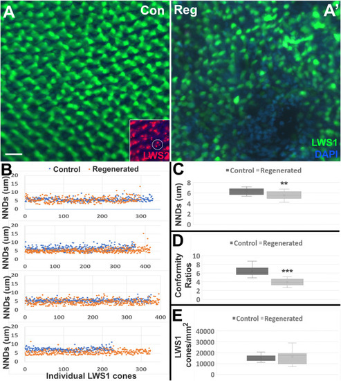
Fig. 2. Localized patterning of LWS1 cones in control and regenerated retinas of adult zebrafish at 21 days following ouabain lesion. A. Representative regions sampled for nearest neighbor distance (NND) analysis from control (Con; A) and contralateral regenerated (Reg; A′) retinas of lws:PAC(H) zebrafish, in which lws1 is reported by GFP (LWS1 cones; green), counterstained with DAPI (blue). Scale bar (applies to A, A′) = 10 μm. Inset in A (same magnification) shows LWS2 cone reporter dsRedExpress forming aggregates in undamaged retina, making the identification of the location of individual LWS2 cones extremely difficult. An example of three aggregates that may or may not be localized to a single cone is encircled. B. Scatterplots show NNDs of individual LWS1 cones in one of three representative regions of four sampled control retinas (blue symbols) and contralateral regenerated retinas (orange symbols). Each graph describes one of the three sampled regions from contralateral retinas. C. Distribution of average NNDs from regions sampled show decreased NNDs of LWS1 cones in regenerated retinas (**p < 0.01; Student's t-test). D. Conformity ratios of the LWS1 pattern are decreased in regenerated retinas (***p < 0.001; Mann-Whitney test), indicating decreased pattern regularity. E. Density of LWS1 cones in the regions sampled are not different in control vs. regenerated retinas. In the boxplots of C-E, the boxes demarcate the 25th and 75th percentiles, the horizontal lines within the boxes designate the medians, the “X” designates the means, and the whiskers represent the upper and lower limits. (For interpretation of the references to color in this figure legend, the reader is referred to the Web version of this article.) |
Reprinted from Experimental Eye Research, 212, Stenkamp, D.L., Viall, D.D., Mitchell, D.M., Evidence of regional specializations in regenerated zebrafish retina, 108789, Copyright (2021) with permission from Elsevier. Full text @ Exp. Eye. Res.