FIGURE SUMMARY
Title

Multiview deconvolution approximation multiphoton microscopy of tissues and zebrafish larvae

Authors
Kapsokalyvas, D., Rosas, R., Janssen, R.W.A., Vanoevelen, J.M., Nabben, M., Strauch, M., Merhof, D., van Zandvoort, M.A.M.J.
Source
Full text @ Sci. Rep.

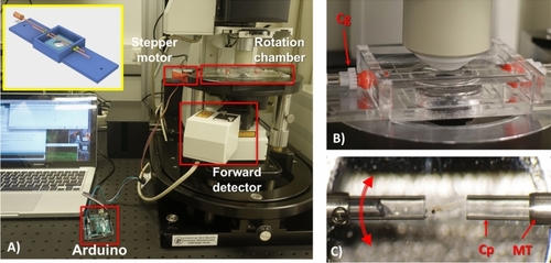
Rotation chamber for Multiview imaging with a two-photon microscope. (A) The rotation chamber mounted on the microscope stage is indicated. The stepper motor is attached to the shaft of the rotation chamber and is controlled by the Arduino board. On the inset of (A) the CAD design of the rotation chamber is shown. (B) The objective is focused on the sample in the rotation chamber. There is ample space for the objective to reach the sample. Below the chamber the condenser lens is positioned. The two cogs responsible for the rotation of the sample are indicated with Cg. (C) An image of the sample when it is ready for imaging. The sample is embedded in the agarose column and is extruded from the capillary allowing the objective to image the sample through the agarose layer. Red arrow indicates rotation direction. MT indicates the metallic tube, Cp indicates the haematocrit glass capillaries.

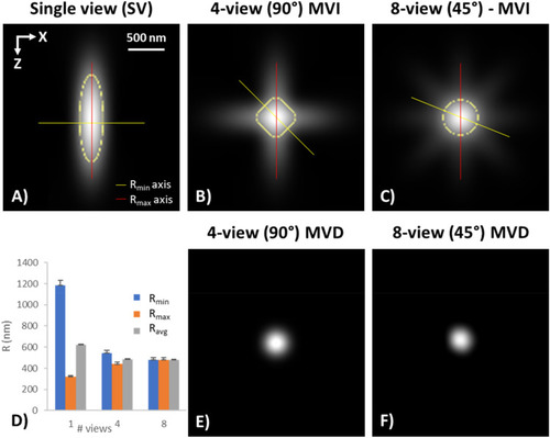
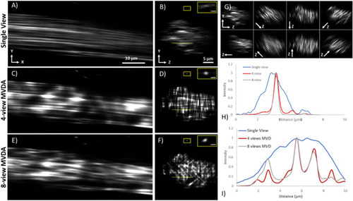
Multiview resolution. (AC) Cross section of the PSF of (A) Single View (SV), (B) 4-views MVI, and (C) 8-views MVI. The yellow line indicates the axis of Rmin, the red line the axis of Rmax, and the yellow dotted line the area of the FWHM from which Ravg is extracted. (D) Graph of the Rmin, Rmax and Ravg of the PSFs in (AC). (E) 4-view deconvolved PSF of 2B. F) 8-view deconvolved PSF of 2C. (Images were created with Fiji26).

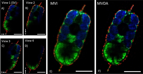
Cross section of the four view Multiview images of a 3dpf zebrafish sample. (AD) Registered individual single view images. Arrow indicates the direction of the z axis of each view. Rotation axis for MVI was on the x-axis which is perpendicular to these images. (E) MVI (fusion) of single images, where the entirety of the sample is visible. (F) MV deconvolution approximation (MVDA) image. Structures appear with better contrast as noise is decreased and resolution improved. Low signal features become better visible in this image. Scale bars 100 μm. (Images were created with Fiji26).

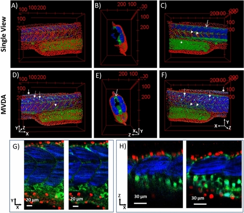
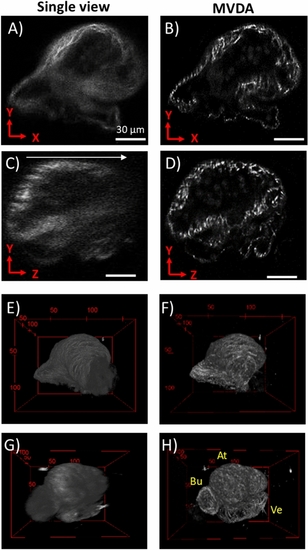
3D images of SV and MVDA. Nuclei in red, autofluorescence and sensory neurons (GFP) in green, muscle fibres and collagen in blue. (A) SV image, (B) SV image rotated 90° to the right. (C) SV rotated 180° compared to (A). (D) MVDA image with the same viewing perspective as A). MVDA image rotated 90° to the right compared to (D). (F) MVDA rotated 180° compared to (D). Arrow heads: myotomes, Diamond arrows collagen in the fins, Open arrows: sensory neurons, Arrows: dendritic like cells. (G) 2D images in the XY plane of SV left and MVDA right. (H) 2D images in the XZ plane of SV left and MVDA right. (Images were created with Fiji26).

Rat-tail tendon imaging based on SHG. Single view (A) XY plane, (B) YZ plane (sagittal section). 4-view Multiview deconvolution (C) XY plane (D) YZ plane (sagittal section). 8-view Multiview deconvolution (E) XY plane (F) YZ plane (sagittal section). (G) YZ planes (sagittal sections) of all individual registered views. The direction of the Z-axis is indicated in each view. (H) Normalized intensity plot profiles of the fibre on the insets of (B), (D), and (F) contrast has been increased for visualization. (I) Normalized intensity plot profiles of the dotted lines in (B), (D), and (F). More individual fibres are revealed in the profiles of 4-view MVDA and 8-view MVDA. Scale bar on the inset of (B), (D), (F) 1 μm. (Images were created with Fiji26).

3D rendering of rat-tail tendon. (A) Single view. (B) 8-view MVD. In the MVD single fibrils are clearly visible. (Images were created with Fiji26).

SHG images of 3-dpf zebrafish heart. (A) Single view YX imaging plane. (B) MVDA YX imaging plane. (C) SV sagittal section (YZ plane), arrow indicates direction of z-stack. (D) MVDA sagittal section. Scale bar: 30 μm. (EH) 3D reconstructions of the zebrafish heart. (E), and (G) SV, (F) and (H) MVDA. (F) and (H) rotated 90° compared to (E) and (G) respectively. At: Atrium, Ve: Ventricle, Bu: Bulbus arteriosus. (Images were created with Fiji26).

Acknowledgments
This image is the copyrighted work of the attributed author or publisher, and ZFIN has permission only to display this image to its users. Additional permissions should be obtained from the applicable author or publisher of the image. Full text @ Sci. Rep.